Pudendal nerve
Main somatic nerve of the perineum (S2–S4), supplying the external anal and urethral sphincters, the perineal muscles and perineal skin. It is blocked at the ischial spine in labor and can be stretched in childbirth or entrapped in cyclists.
Core anatomy
- Location
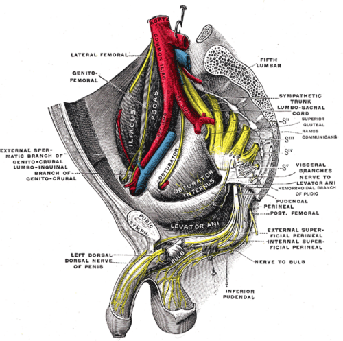
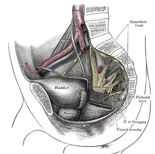
Leaves the pelvis, curves around the ischial spine, and runs forward in the lateral wall of the ischioanal fossa to the perineum.
- Root values
Ventral rami of S2, S3 and S4 (from the sacral plexus).
- Origin
Sacral plexus, on the posterior pelvic wall.
- Course
Leaves the pelvis through the greater sciatic foramen below piriformis, crosses the back of the sacrospinous ligament just medial to the ischial spine (medial to the internal pudendal vessels), and re-enters the perineum through the lesser sciatic foramen. It then runs in the pudendal (Alcock) canal, a split in the obturator internus fascia, with the internal pudendal vessels.
- Branches
- Inferior rectal (anal) nerve: crosses the ischioanal fossa to the external anal sphincter, the lower anal canal (below the pectinate line) and perianal skin
- Perineal nerve: superficial branches (posterior scrotal/labial nerves) to the skin; deep branches to bulbospongiosus, ischiocavernosus, the transverse perineal muscles and the external urethral sphincter
- Dorsal nerve of the penis or clitoris: sensory to the glans and most of the shaft
- Motor
External anal sphincter, external urethral sphincter, perineal muscles, and part of levator ani.
- Sensory
Most of the perineal skin, the posterior scrotum or labia, the penis or clitoris, the lower quarter of the vagina, and the anal canal below the pectinate line.
- Relations
The internal pudendal vessels run with it throughout. The anterior perineum (mons, anterior scrotum/labia) is supplied instead by the ilioinguinal and genitofemoral nerves, and the lateral perineum by the posterior femoral cutaneous nerve.
- Notes
"S2, 3, 4 keep the poop off the floor" (and the pee): pudendal and pelvic splanchnic nerves serve continence.
Muscles innervated
- Pelvic diaphragm — Nerve to levator ani (direct branches of the ventral rami of S3–S4), with contributions from the inferior rectal and perineal branches of the pudendal nerve. Coccygeus: S3–S4 ventral rami.
Other structures innervated
Found in
Mentioned in
Generated automatically from links in other entries.
Swipe sideways to see the whole diagram →


Clinical correlations
Pudendal nerve block PANCE
- Mechanism / lesion
Local anesthetic injected transvaginally through the sacrospinous ligament just medial and posterior to the ischial spine, where the nerve crosses. Aspirate first to avoid the internal pudendal vessels.
- Classic presentation
Anesthetizes most of the perineum for the second stage of labor, forceps delivery or perineal repair. The anterior perineum (ilioinguinal/genitofemoral) and lateral perineum (posterior femoral cutaneous) still need local infiltration.
Obstetric pudendal neuropathy
- Mechanism / lesion
Stretch injury during prolonged second stage or instrumental delivery.
- Classic presentation
Fecal and/or urinary incontinence with reduced perineal sensation; usually improves over months.
Pudendal neuralgia (entrapment)
- Mechanism / lesion
Compression between the sacrotuberous and sacrospinous ligaments or in the pudendal canal; associated with prolonged cycling and pelvic surgery.
- Classic presentation
Burning pain in the perineum, genitals or rectum that is worse sitting and relieved by standing, lying down or sitting on a toilet seat; no objective sensory loss (Nantes criteria). A diagnostic pudendal block helps confirm it.
High-yield PANCE pearls
- Pudendal = S2–S4; supplies the external anal and urethral sphincters and perineal skin.
- Pudendal block: needle just medial to the ischial spine through the sacrospinous ligament.
- Perineal pain worse with sitting in a cyclist → pudendal neuralgia.
- Saddle anesthesia + loss of anal tone → cauda equina (S2–S5) — not an isolated pudendal problem.
Imaging anatomy
- CT
CT (or ultrasound/fluoroscopy) guides pudendal nerve blocks at the ischial spine or in the pudendal canal.
- MRI
MR neurography can show the nerve and sites of compression; MRI excludes masses along its course.
Physical exam
- Surface landmarks
The ischial spine, palpable transvaginally or transrectally at about 4 and 8 o'clock, marks where the nerve crosses the sacrospinous ligament.
- Palpation
Tenderness over the ischial spine on internal exam supports pudendal entrapment.
- Strength / function testing
Perianal and perineal pinprick and light touch (S3–S5 "saddle"), anal wink (anocutaneous reflex), bulbocavernosus reflex and DRE for resting tone and voluntary anal contraction (a key part of the ASIA exam in spinal cord injury).
Self-quiz
During a transvaginal pudendal block for a forceps delivery, local anesthetic is injected just medial to the ischial spine. After the block the patient still feels pain at the anterior labia. Which nerve supplies that area?
Correct answer: B
The anterior labia (and mons) are supplied by the ilioinguinal nerve and the genital branch of the genitofemoral nerve (L1), which a pudendal block does not reach, so local infiltration is added.
Show answer
B
The anterior labia (and mons) are supplied by the ilioinguinal nerve and the genital branch of the genitofemoral nerve (L1), which a pudendal block does not reach, so local infiltration is added.
Which muscle is supplied by the inferior rectal branch of the pudendal nerve?
Correct answer: B
The inferior rectal nerve supplies the voluntary external anal sphincter and perianal skin. The internal sphincter is smooth muscle under autonomic control; bulbospongiosus is supplied by the perineal nerve.
Show answer
B
The inferior rectal nerve supplies the voluntary external anal sphincter and perianal skin. The internal sphincter is smooth muscle under autonomic control; bulbospongiosus is supplied by the perineal nerve.
The pudendal nerve re-enters the perineum through which opening?
Correct answer: C
The pudendal nerve leaves the pelvis through the greater sciatic foramen below piriformis, curves around the ischial spine, and enters the perineum through the lesser sciatic foramen into the pudendal canal.
Show answer
C
The pudendal nerve leaves the pelvis through the greater sciatic foramen below piriformis, curves around the ischial spine, and enters the perineum through the lesser sciatic foramen into the pudendal canal.
A 45-year-old competitive cyclist has burning perineal pain that is worse when sitting and relieved by standing or sitting on a toilet seat. Sensation is normal on exam. What is the most likely diagnosis?
Correct answer: E
Pudendal neuralgia (entrapment between the sacrotuberous and sacrospinous ligaments or in the pudendal canal) causes burning perineal, genital or rectal pain that is worse sitting, with no objective sensory loss. It is associated with prolonged cycling. A diagnostic pudendal block helps confirm it.
Show answer
E
Pudendal neuralgia (entrapment between the sacrotuberous and sacrospinous ligaments or in the pudendal canal) causes burning perineal, genital or rectal pain that is worse sitting, with no objective sensory loss. It is associated with prolonged cycling. A diagnostic pudendal block helps confirm it.
What forms the pudendal (Alcock) canal?
Correct answer: B
The pudendal canal is a split in the obturator internus fascia on the lateral wall of the ischioanal fossa. The pudendal nerve runs in it with the internal pudendal vessels after re-entering through the lesser sciatic foramen.
Show answer
B
The pudendal canal is a split in the obturator internus fascia on the lateral wall of the ischioanal fossa. The pudendal nerve runs in it with the internal pudendal vessels after re-entering through the lesser sciatic foramen.
Flashcards
See also
- Sacral plexus
- Perineum
- Ischioanal fossa
- Anal canal
- Lower limb nerve injuries (comparison table)
- Anal canal above vs below the pectinate line (comparison table)