Compartments of the leg
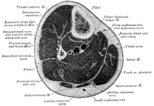
Four tight osteofascial compartments — anterior, lateral, superficial posterior and deep posterior — each with its own muscles, nerve and vessels. Rising pressure after fractures or crush injuries causes compartment syndrome, a surgical emergency.
Core anatomy
- Location
The leg between the knee and ankle, enclosed by the deep (crural) fascia.
- Boundaries
- anterior compartment
- Tibia (medially), interosseous membrane (posteriorly), fibula and anterior intermuscular septum (laterally), deep fascia (anteriorly)
- lateral compartment
- Fibula (medially), anterior and posterior intermuscular septa, deep fascia
- superficial posterior compartment
- Transverse intermuscular septum (deep) and deep fascia
- deep posterior compartment
- Tibia, interosseous membrane and fibula (anteriorly), transverse intermuscular septum (posteriorly)
- Contents
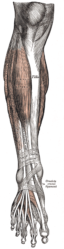
- Anterior: tibialis anterior, extensor hallucis longus, extensor digitorum longus, fibularis tertius — dorsiflexion and toe extension; deep fibular nerve; anterior tibial artery
- Lateral: fibularis longus and brevis — eversion; superficial fibular nerve (no major artery; supplied by fibular artery branches)
- Superficial posterior: gastrocnemius, soleus, plantaris (triceps surae and calcaneal tendon) — plantarflexion; branches of the tibial nerve; sural nerve and small saphenous vein lie superficial to it
- Deep posterior: tibialis posterior, flexor digitorum longus, flexor hallucis longus (and popliteus) — inversion, toe flexion; tibial nerve; posterior tibial and fibular arteries
- Communications
The compartments are closed spaces bounded by bone and non-expansile fascia — the reason pressure rises quickly when they swell.
Mentioned in
Generated automatically from links in other entries.
Swipe sideways to see the whole diagram →


Clinical correlations
Acute compartment syndrome PANCE
- Mechanism / lesion
Raised pressure within a closed compartment reduces perfusion → muscle and nerve ischemia. Causes: tibial fractures (most common), crush injury, reperfusion after arterial repair, tight casts, burns, intramuscular bleeding. The anterior compartment is most often affected.
- Classic presentation
Pain out of proportion to the injury and pain on passive stretch of the compartment's muscles (earliest signs); tense compartment; paresthesias (first web space for the anterior compartment). Pulselessness and paralysis are late — pulses are usually present.
- Notes
Clinical diagnosis; measure compartment pressure if uncertain (fasciotomy if delta pressure — diastolic BP minus compartment pressure — is below 30 mm Hg). Treatment: remove casts/dressings, keep the limb at heart level, and emergency four-compartment fasciotomy.
Chronic exertional compartment syndrome
- Mechanism / lesion
Reversible pressure rise during exercise, usually the anterior or lateral compartment, in runners.
- Classic presentation
Predictable aching or tightness after a set distance, relieved by rest within minutes; normal exam at rest. Diagnosed with pre- and post-exercise pressures.
High-yield PANCE pearls
- Compartment syndrome: pain out of proportion + pain on passive stretch → fasciotomy; pulses are often present.
- Delta pressure (diastolic − compartment) < 30 mm Hg → fasciotomy.
- Anterior compartment = deep fibular nerve + anterior tibial artery (dorsiflexion); lateral = superficial fibular (eversion).
- Deep posterior = tibial nerve + posterior tibial and fibular arteries (inversion, toe flexion).
Imaging anatomy
- X-ray
Identifies the underlying fracture — imaging does not diagnose compartment syndrome.
- MRI
Edema of the involved compartment in chronic exertional compartment syndrome (not needed for acute cases).
Physical exam
- Surface landmarks
The anterior compartment lies just lateral to the subcutaneous tibial border; the lateral compartment over the fibula; the posterior compartments form the calf.
- Palpation
Palpate each compartment for tense, "woody" firmness.
- Strength / function testing
Passive stretch of each compartment: plantarflex the toes/ankle (anterior), invert the foot (lateral), dorsiflex the ankle (superficial posterior), extend the toes (deep posterior). Check sensation in the first web space (deep fibular), dorsum (superficial fibular) and sole (tibial). Measure compartment pressures if the exam is unreliable (e.g. obtunded patient).
Self-quiz
Four hours after casting of a tibial shaft fracture, a 22-year-old has severe leg pain not relieved by morphine, worsened by passive plantarflexion of the toes, and numbness between the first and second toes. The dorsalis pedis pulse is palpable. What is the most appropriate next step?
Correct answer: C
Anterior compartment syndrome: pain out of proportion, pain with passive stretch of the anterior compartment muscles, and deep fibular nerve numbness (first web space). A palpable pulse does not exclude it. Release all constrictive dressings and perform emergency fasciotomy; keep the limb at heart level, not elevated.
Show answer
C
Anterior compartment syndrome: pain out of proportion, pain with passive stretch of the anterior compartment muscles, and deep fibular nerve numbness (first web space). A palpable pulse does not exclude it. Release all constrictive dressings and perform emergency fasciotomy; keep the limb at heart level, not elevated.
Which compartment of the leg contains the posterior tibial artery and tibial nerve?
Correct answer: D
The deep posterior compartment contains tibialis posterior, FDL, FHL, the tibial nerve, and the posterior tibial and fibular arteries.
Show answer
D
The deep posterior compartment contains tibialis posterior, FDL, FHL, the tibial nerve, and the posterior tibial and fibular arteries.
Which nerve supplies the muscles of the lateral compartment of the leg, and what movement do they produce?
Correct answer: C
The lateral compartment (fibularis longus and brevis) is supplied by the superficial fibular nerve and everts the foot. It has no major artery of its own; it is supplied by branches of the fibular artery.
Show answer
C
The lateral compartment (fibularis longus and brevis) is supplied by the superficial fibular nerve and everts the foot. It has no major artery of its own; it is supplied by branches of the fibular artery.
Which compartment pressure measurement supports emergency fasciotomy?
Correct answer: B
Compartment syndrome is a clinical diagnosis, but when pressures are measured, a delta pressure (diastolic BP − compartment pressure) below 30 mm Hg supports fasciotomy.
Show answer
B
Compartment syndrome is a clinical diagnosis, but when pressures are measured, a delta pressure (diastolic BP − compartment pressure) below 30 mm Hg supports fasciotomy.
A 24-year-old runner has predictable tightness and aching in the front of both legs after about 3 km, relieved within minutes of stopping. The exam at rest is normal. What is the most likely diagnosis?
Correct answer: D
Chronic exertional compartment syndrome is a reversible pressure rise during exercise, usually in the anterior or lateral compartment, with symptoms at a predictable distance that settle with rest. It is diagnosed with pre- and post-exercise compartment pressures.
Show answer
D
Chronic exertional compartment syndrome is a reversible pressure rise during exercise, usually in the anterior or lateral compartment, with symptoms at a predictable distance that settle with rest. It is diagnosed with pre- and post-exercise compartment pressures.
Flashcards
See also
- Tibia
- Fibula
- Common fibular nerve
- Tibial nerve
- Triceps surae and calcaneal tendon
- Lower limb nerve injuries (comparison table)
- Severe pain under a new cast (clinical case)