Superior mesenteric artery
Artery of the midgut, arising from the aorta at L1 behind the pancreas. Its occlusion causes acute mesenteric ischemia, and its angle with the aorta explains SMA and nutcracker syndromes.
Core anatomy
- Location
Arises about 1 cm below the celiac trunk at about L1, behind the neck of the pancreas.
- Origin
- Course
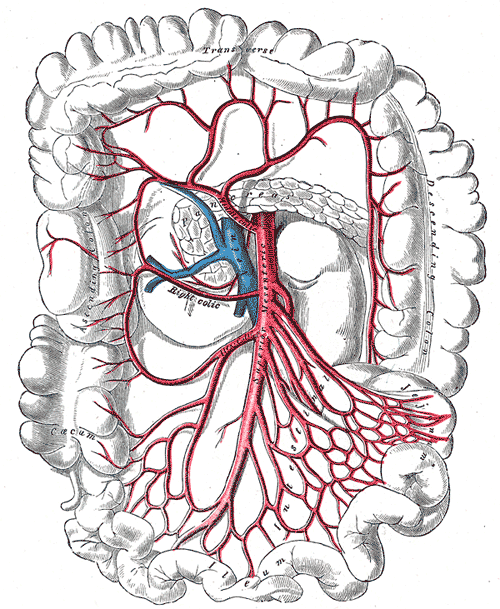
Descends anterior to the left renal vein, the uncinate process of the pancreas, and the 3rd part of the duodenum, then enters the root of the mesentery toward the right iliac fossa.
- Branches
- Inferior pancreaticoduodenal arteries
- Jejunal and ileal arteries (from its left side), forming arcades and vasa recta
- Middle colic artery (transverse colon)
- Right colic artery (ascending colon)
- Ileocolic artery (terminal ileum, cecum, appendix via the appendicular artery)
- Supplies
Midgut: duodenum distal to the major papilla, jejunum, ileum, cecum, appendix, ascending colon, and the proximal two-thirds of the transverse colon.
- Anastomoses
Pancreaticoduodenal arcades with the celiac trunk; the marginal artery and middle colic–left colic connection with the inferior mesenteric artery.
- Relations
The left renal vein passes between the SMA and the aorta; the 3rd part of the duodenum also passes between them. The superior mesenteric vein lies to its right.
Structures supplied
Mentioned in
Generated automatically from links in other entries.
Swipe sideways to see the whole diagram →

Clinical correlations
Acute mesenteric ischemia PANCE
- Mechanism / lesion
Embolism (often from atrial fibrillation or recent MI; typically lodges a few cm beyond the origin, sparing the proximal jejunum), thrombosis of an atherosclerotic origin, non-occlusive ischemia (low-flow states), or mesenteric venous thrombosis.
- Classic presentation
Sudden severe abdominal pain out of proportion to the exam, nausea, vomiting, possibly bloody stool. Lactate rises late. CT angiography; treatment is emergency revascularization and resection of dead bowel.
Chronic mesenteric ischemia PANCE
- Mechanism / lesion
Atherosclerotic stenosis of usually two or more mesenteric arteries.
- Classic presentation
"Intestinal angina": postprandial pain 15–60 minutes after eating, food fear, and weight loss, in a patient with vascular disease.
SMA syndrome and nutcracker syndrome
- Mechanism / lesion
A narrowed aortomesenteric angle compresses the 3rd part of the duodenum (SMA syndrome) or the left renal vein (nutcracker syndrome).
- Classic presentation
SMA syndrome causes postprandial pain and vomiting after rapid weight loss. Nutcracker causes hematuria, left flank pain and a left varicocele.
High-yield PANCE pearls
- SMA (L1) = midgut: distal duodenum → proximal 2/3 transverse colon.
- Pain out of proportion to exam + AF → SMA embolus → CT angiography.
- Chronic mesenteric ischemia: postprandial pain + food fear + weight loss.
- Left renal vein and 3rd part of duodenum pass between SMA and aorta.
Imaging anatomy
- X-ray
Late signs of ischemia (pneumatosis intestinalis, portal venous gas, thumbprinting). Often normal early.
- CT
CT angiography is the test of choice for acute mesenteric ischemia (filling defect, bowel wall changes, pneumatosis). Sagittal images show the SMA origin and the aortomesenteric angle.
- Ultrasound
Duplex screens for SMA stenosis (elevated peak systolic velocity) in chronic mesenteric ischemia; limited by bowel gas.
- Key views & landmarks
- CT angiography (arterial phase, sagittal reformats)
Physical exam
- Palpation
A soft abdomen with minimal tenderness despite severe pain is the classic early finding in acute mesenteric ischemia. Peritoneal signs appear late (infarction).
- Strength / function testing
Listen for an epigastric bruit. Look for atrial fibrillation (irregularly irregular pulse).
Self-quiz
The SMA supplies which part of the colon?
Correct answer: C
The SMA supplies the midgut, including the cecum, ascending colon and proximal two-thirds of the transverse colon (via the ileocolic, right colic and middle colic arteries).
Show answer
C
The SMA supplies the midgut, including the cecum, ascending colon and proximal two-thirds of the transverse colon (via the ileocolic, right colic and middle colic arteries).
A 64-year-old woman with vascular disease has abdominal pain 30 minutes after meals, has lost 9 kg, and now avoids eating. What is the most likely diagnosis?
Correct answer: B
Intestinal angina: postprandial pain, food fear and weight loss from atherosclerotic mesenteric stenosis (chronic mesenteric ischemia).
Show answer
B
Intestinal angina: postprandial pain, food fear and weight loss from atherosclerotic mesenteric stenosis (chronic mesenteric ischemia).
Which vessel passes between the SMA and the abdominal aorta?
Correct answer: B
The left renal vein crosses anterior to the aorta beneath the SMA. Compression here (nutcracker syndrome) can cause hematuria and a left varicocele.
Show answer
B
The left renal vein crosses anterior to the aorta beneath the SMA. Compression here (nutcracker syndrome) can cause hematuria and a left varicocele.
A thin young man has hematuria, left flank pain and a left varicocele. Imaging shows compression of the left renal vein. Between which structures is it compressed?
Correct answer: E
In nutcracker syndrome, a narrow aortomesenteric angle compresses the left renal vein between the SMA and aorta, causing hematuria, left flank pain and a left varicocele. The same angle compresses the duodenum in SMA syndrome.
Show answer
E
In nutcracker syndrome, a narrow aortomesenteric angle compresses the left renal vein between the SMA and aorta, causing hematuria, left flank pain and a left varicocele. The same angle compresses the duodenum in SMA syndrome.
Which branch of the SMA supplies the appendix?
Correct answer: B
The appendicular artery is a branch of the ileocolic artery, which also supplies the terminal ileum and cecum. The left colic artery belongs to the IMA.
Show answer
B
The appendicular artery is a branch of the ileocolic artery, which also supplies the terminal ileum and cecum. The left colic artery belongs to the IMA.
Flashcards
See also
- Small intestine (jejunum and ileum)
- Colon
- Abdominal aorta
- Duodenum
- Foregut, midgut and hindgut (comparison table)
- Pain that moves to the right lower quadrant (clinical case)