Thymus
Primary lymphoid organ in the anterior and superior mediastinum where T cells mature. It is large in children, involutes after puberty, and thymoma is associated with myasthenia gravis.
Core anatomy
- Location
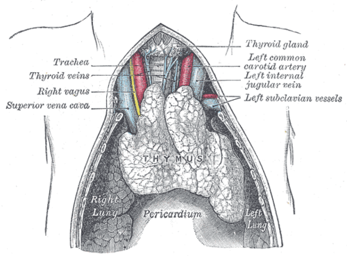
Superior and anterior mediastinum, behind the manubrium and in front of the great vessels and pericardium. It may extend into the neck.
- Parts
- Two lobes, each with an outer cortex (immature T cells) and inner medulla (mature T cells, Hassall corpuscles)
- Relatively large in infancy; replaced progressively by fat after puberty (involution)
- Relations
Posterior to the sternum (manubrium); anterior to the left brachiocephalic vein, the arch of the aorta and its branches, and the pericardium.
- Arterial supply
Branches of the internal thoracic artery and inferior thyroid arteries.
- Venous drainage
Left brachiocephalic, internal thoracic and inferior thyroid veins.
- Notes
Develops from the third pharyngeal pouch (endoderm), together with the inferior parathyroid glands.
Found in
Mentioned in
Generated automatically from links in other entries.
Swipe sideways to see the whole diagram →

Clinical correlations
Thymoma and myasthenia gravis PANCE
- Mechanism / lesion
Thymic epithelial tumor. A substantial proportion of thymoma patients have myasthenia gravis, and a minority of myasthenia patients have a thymoma; thymic hyperplasia is more common in MG. Other associations: pure red cell aplasia, hypogammaglobulinemia. verify
- Classic presentation
Anterior mediastinal mass (often incidental), or symptoms of MG: fatigable weakness, ptosis, diplopia, bulbar symptoms. All MG patients should have chest CT to look for thymoma. Thymectomy benefits selected patients.
DiGeorge syndrome (22q11.2 deletion) PANCE
- Mechanism / lesion
Failure of development of the 3rd and 4th pharyngeal pouches.
- Classic presentation
Thymic aplasia (T-cell deficiency, recurrent viral/fungal infections), hypocalcemia (absent parathyroids, tetany), conotruncal heart defects (tetralogy of Fallot, truncus arteriosus), and facial anomalies. The thymic shadow is absent on neonatal CXR.
High-yield PANCE pearls
- Anterior mediastinal mass: 4 T's: Thymoma, Teratoma (germ cell), Thyroid (retrosternal goiter), Terrible lymphoma.
- Myasthenia gravis → CT chest for thymoma.
- DiGeorge: absent thymus (no thymic shadow), hypocalcemia, conotruncal heart defects; 22q11.2 deletion.
- The thymus is normally prominent on infant CXR (sail sign), not a mass.
Imaging anatomy
- X-ray
In infants the normal thymus can look like a mediastinal mass; the "sail sign" (a triangular thymic shadow) is normal. The thymic shadow is absent in DiGeorge syndrome. In adults, anterior mediastinal masses are seen on the lateral film as retrosternal opacities.
- CT
CT chest is the main test for thymoma (a well-defined anterior mediastinal mass) and for distinguishing it from lymphoma, germ cell tumors and goiter.
- MRI
Chemical-shift MRI helps distinguish thymic hyperplasia (which contains fat and loses signal) from thymoma.
- Key views & landmarks
- CT chest with contrast (anterior mediastinum)
Physical exam
- Strength / function testing
For MG: sustained upgaze (fatigable ptosis), the ice pack test (ptosis improves with cold), and repetitive testing of proximal muscle strength.
Self-quiz
A 45-year-old woman has double vision and drooping eyelids that worsen at the end of the day. Acetylcholine receptor antibodies are positive. Which imaging study is indicated?
Correct answer: B
Every patient with myasthenia gravis needs chest CT (or MRI) to look for a thymoma in the anterior mediastinum. Thymectomy is indicated for thymoma and can benefit selected patients without one.
Show answer
B
Every patient with myasthenia gravis needs chest CT (or MRI) to look for a thymoma in the anterior mediastinum. Thymectomy is indicated for thymoma and can benefit selected patients without one.
A newborn has tetany, recurrent thrush, and a conotruncal heart defect. The chest radiograph shows no thymic shadow. Which embryologic structures failed to develop?
Correct answer: B
DiGeorge syndrome (22q11.2 deletion): the 3rd and 4th pharyngeal pouches fail to develop, so the thymus (T cells) and parathyroids (calcium) are absent, with conotruncal defects.
Show answer
B
DiGeorge syndrome (22q11.2 deletion): the 3rd and 4th pharyngeal pouches fail to develop, so the thymus (T cells) and parathyroids (calcium) are absent, with conotruncal defects.
Which is NOT one of the classic causes of an anterior mediastinal mass?
Correct answer: E
Neurogenic tumors arise from the sympathetic chain and nerve roots in the posterior mediastinum. The anterior "4 T's" are thymoma, teratoma, thyroid, and "terrible" lymphoma.
Show answer
E
Neurogenic tumors arise from the sympathetic chain and nerve roots in the posterior mediastinum. The anterior "4 T's" are thymoma, teratoma, thyroid, and "terrible" lymphoma.
The thymus develops from which embryologic structure?
Correct answer: B
The thymus develops from the third pharyngeal pouch (endoderm), together with the inferior parathyroid glands. DiGeorge syndrome reflects failure of the 3rd and 4th pouches.
Show answer
B
The thymus develops from the third pharyngeal pouch (endoderm), together with the inferior parathyroid glands. DiGeorge syndrome reflects failure of the 3rd and 4th pouches.
A chest radiograph of a healthy 6-month-old shows a triangular soft-tissue density extending from the upper mediastinum. What is it most likely to be?
Correct answer: E
The thymus is relatively large in infancy and normally visible on CXR as the "sail sign"; it isn't a mass. It involutes after puberty and is replaced by fat.
Show answer
E
The thymus is relatively large in infancy and normally visible on CXR as the "sail sign"; it isn't a mass. It involutes after puberty and is replaced by fat.