Azygos vein
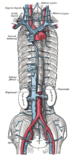
Vein on the right side of the posterior mediastinum that drains the posterior thoracic wall. It arches over the right lung root into the SVC and is a key collateral pathway between the caval systems.
Core anatomy
- Location
Posterior mediastinum, on the right side of the thoracic vertebral bodies.
- Formation
Usually from the right ascending lumbar and right subcostal veins, and sometimes a connection with the IVC, entering the thorax through the aortic hiatus or the right crus of the diaphragm. verify
- Course
Ascends on the right of the vertebral bodies (with the thoracic duct and thoracic aorta to its left). At about T4 it arches anteriorly over the root of the right lung (right main bronchus) to join the posterior superior vena cava.
- Tributaries
- Right posterior intercostal veins (4th–11th) and the right superior intercostal vein
- Hemiazygos vein (lower left posterior thorax) and accessory hemiazygos vein (upper left), which cross to the right behind the aorta
- Esophageal, bronchial, mediastinal and pericardial veins
- Drains into
- Relations
The arch of the azygos vein is on the right of the trachea and esophagus above the right main bronchus. It lies behind the esophagus lower down.
Tributaries
Found in
Mentioned in
- Esophagus
- Intercostal space
- Lungs
- Main bronchi
- Sternum
- Superior vena cava
- Thoracic aorta
- Thoracic duct
- Trachea
Generated automatically from links in other entries.

Clinical correlations
Collateral pathway in caval obstruction PANCE
- Mechanism / lesion
The azygos system connects the SVC and IVC via the lumbar veins.
- Classic presentation
SVC obstruction below the azygos entry: blood returns from the upper body through the azygos in reverse. IVC obstruction: lower-body blood returns through the lumbar–azygos route. The azygos vein becomes dilated on imaging.
Esophageal varices (systemic side) PANCE
- Mechanism / lesion
In portal hypertension, portal blood reaches the systemic circulation via the left gastric vein → esophageal veins → azygos vein.
- Classic presentation
Dilated submucosal veins of the distal esophagus that can bleed massively.
Azygos lobe
- Mechanism / lesion
A normal variant in which the azygos vein passes through the apical right upper lobe, carrying a fold of pleura with it (the azygos fissure).
- Classic presentation
A curved thin line in the right upper zone on CXR ending in a teardrop density. Clinically insignificant.
High-yield PANCE pearls
- Azygos arches over the right main bronchus at T4 into the SVC.
- Azygos = SVC–IVC collateral; dilated with caval obstruction or azygos continuation of the IVC.
- Esophageal varices: left gastric (portal) ↔ esophageal veins → azygos (systemic).
Imaging anatomy
- X-ray
The azygos arch appears as a small oval opacity in the right tracheobronchial angle; enlargement suggests raised central venous pressure, caval obstruction, or azygos continuation. Azygos fissure (variant).
- CT
Shows the course, dilation (azygos continuation of an interrupted IVC, SVC obstruction) and collaterals.
- Key views & landmarks
- CT chest with contrast
Physical exam
- Strength / function testing
Not examined directly. Look for chest and abdominal wall collateral veins in caval obstruction.
Self-quiz
At what level does the azygos vein arch over the root of the right lung to join the SVC?
Correct answer: B
The azygos vein arches over the right lung root at about T4, the level of the sternal angle, entering the posterior SVC.
Show answer
B
The azygos vein arches over the right lung root at about T4, the level of the sternal angle, entering the posterior SVC.
In a patient with obstruction of the inferior vena cava, which vessel is a major collateral route for venous return from the lower body to the heart?
Correct answer: B
The azygos/hemiazygos system connects lumbar veins (IVC territory) to the SVC, providing collateral return when the IVC is blocked.
Show answer
B
The azygos/hemiazygos system connects lumbar veins (IVC territory) to the SVC, providing collateral return when the IVC is blocked.
Which vessels drain the posterior intercostal spaces of the left lower thorax before joining the azygos vein?
Correct answer: B
The hemiazygos vein drains the lower left posterior intercostal veins and crosses behind the aorta to join the azygos vein (the accessory hemiazygos drains the upper left spaces).
Show answer
B
The hemiazygos vein drains the lower left posterior intercostal veins and crosses behind the aorta to join the azygos vein (the accessory hemiazygos drains the upper left spaces).