Vermiform appendix
Blind-ending tube arising from the cecum where the teniae coli converge. Acute appendicitis is the most common abdominal surgical emergency.
Core anatomy
- Location
Right iliac fossa. Its base is on the posteromedial cecum about 2 cm below the ileocecal valve, where the three teniae coli converge (a surgical guide to finding it). The tip is variable (most often retrocecal, also pelvic, subcecal, pre- or post-ileal).
- Parts
- Base, body and tip; lumen rich in lymphoid tissue
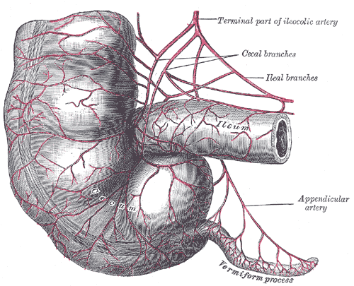
- Mesoappendix: a peritoneal fold from the terminal ileum mesentery, containing the appendicular artery
- Relations
The base projects to McBurney point, one-third of the way from the right ASIS to the umbilicus. A retrocecal appendix lies against the psoas; a pelvic appendix against the obturator internus, bladder or rectum.
- Arterial supply
Appendicular artery, a branch of the ileocolic artery (from the superior mesenteric artery).
- Venous drainage
Appendicular vein → ileocolic vein → SMV → portal vein (portal pyelophlebitis and liver abscess are rare complications).
- Innervation
Visceral afferents with sympathetic fibers to about T10, so early pain refers to the periumbilical region.
Swipe sideways to see the whole diagram →

Clinical correlations
Acute appendicitis PANCE
- Mechanism / lesion
Luminal obstruction (fecalith, lymphoid hyperplasia, rarely tumor) → distension, ischemia, bacterial invasion, and perforation if untreated.
- Classic presentation
Periumbilical pain migrating to the RLQ, then anorexia, nausea/vomiting (usually after the pain begins), and low-grade fever. McBurney point tenderness, rebound and guarding. Signs by position: Rovsing, psoas (retrocecal), obturator (pelvic). Leukocytosis. Presentation can be atypical in children, older adults and pregnancy (the appendix is displaced upward).
Appendiceal neoplasms
- Mechanism / lesion
Neuroendocrine (carcinoid) tumors are among the most common appendiceal tumors, often found incidentally after appendectomy; also mucinous neoplasms.
- Classic presentation
Usually incidental. Mucinous tumors can cause pseudomyxoma peritonei.
High-yield PANCE pearls
- Appendicitis sequence: periumbilical pain → RLQ, anorexia, then vomiting.
- Signs: McBurney, Rovsing (LLQ pressure → RLQ pain), psoas (retrocecal), obturator (pelvic).
- Imaging: CT in adults; ultrasound first in children and pregnancy (MRI if US non-diagnostic in pregnancy).
- Alvarado score helps risk-stratify.
Imaging anatomy
- X-ray
Rarely helpful; an appendicolith may be visible.
- CT
CT with IV contrast is the most accurate test in adults: a dilated appendix (more than about 6 mm), wall thickening and enhancement, periappendiceal fat stranding, appendicolith, abscess or perforation. verify
- MRI
Preferred in pregnancy when ultrasound is non-diagnostic (no radiation).
- Ultrasound
First-line in children and pregnant patients (graded compression): a non-compressible, blind-ending tubular structure larger than about 6 mm in diameter, a target appearance in cross-section, periappendiceal fluid, and focal tenderness.
- Key views & landmarks
- Graded-compression ultrasound of the RLQ
- CT abdomen/pelvis with IV contrast
Physical exam
- Surface landmarks
McBurney point: one-third of the distance from the right ASIS to the umbilicus.
- Palpation
Maximal tenderness at McBurney point; rebound tenderness and guarding with peritoneal involvement.
Rovsing sign
- Technique
Press deeply in the left lower quadrant.
- Positive test
Pain felt in the right lower quadrant.
- Significance
Appendicitis (peritoneal irritation in the RLQ).
Psoas sign
- Technique
Patient lies on the left side; the examiner passively extends the right hip (or the supine patient flexes the right hip against resistance).
- Positive test
RLQ pain.
- Significance
Inflamed appendix against the psoas, typical of a retrocecal appendix. Also positive with psoas abscess.
Obturator sign
- Technique
With the patient supine, flex the right hip and knee to 90° and passively internally rotate the hip.
- Positive test
Hypogastric or RLQ pain.
- Significance
Inflammation adjacent to obturator internus, typical of a pelvic appendix.
Self-quiz
A 21-year-old woman has 18 hours of pain that began around the umbilicus and moved to the right lower quadrant, with anorexia and one episode of vomiting. Pressing in the left lower quadrant causes pain in the right lower quadrant. What is this sign called?
Correct answer: B
Rovsing sign (LLQ pressure producing RLQ pain) supports appendicitis. Murphy sign is for cholecystitis; psoas and obturator signs suggest retrocecal and pelvic positions respectively.
Show answer
B
Rovsing sign (LLQ pressure producing RLQ pain) supports appendicitis. Murphy sign is for cholecystitis; psoas and obturator signs suggest retrocecal and pelvic positions respectively.
A 24-year-old pregnant woman at 20 weeks has right-sided abdominal pain and suspected appendicitis. What is the first imaging test?
Correct answer: B
In pregnancy (and children), ultrasound is first. If it's non-diagnostic, MRI is preferred over CT to avoid radiation.
Show answer
B
In pregnancy (and children), ultrasound is first. If it's non-diagnostic, MRI is preferred over CT to avoid radiation.
Why does early appendicitis cause periumbilical pain?
Correct answer: B
Visceral afferents from the appendix (midgut) enter at about T10, the dermatome of the umbilicus, so early pain is referred there before the parietal peritoneum is involved.
Show answer
B
Visceral afferents from the appendix (midgut) enter at about T10, the dermatome of the umbilicus, so early pain is referred there before the parietal peritoneum is involved.
A surgeon has difficulty finding the appendix. Which feature reliably leads to its base?
Correct answer: B
The three teniae coli converge at the base of the appendix on the cecum, a reliable intraoperative guide.
Show answer
B
The three teniae coli converge at the base of the appendix on the cecum, a reliable intraoperative guide.
A patient with appendicitis has right lower quadrant pain when the examiner passively extends the right hip. Where is the appendix most likely positioned?
Correct answer: C
A positive psoas sign (pain on hip extension) suggests an inflamed retrocecal appendix lying against the psoas. A positive obturator sign (pain on internal rotation of the flexed hip) suggests a pelvic appendix against obturator internus.
Show answer
C
A positive psoas sign (pain on hip extension) suggests an inflamed retrocecal appendix lying against the psoas. A positive obturator sign (pain on internal rotation of the flexed hip) suggests a pelvic appendix against obturator internus.
Flashcards
See also
- Small intestine (jejunum and ileum)
- Colon
- Psoas major
- Foregut, midgut and hindgut (comparison table)
- Pain that moves to the right lower quadrant (clinical case)