Pronator teres
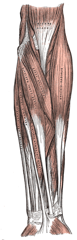
Superficial muscle of the anterior forearm. It pronates the forearm, forms the medial border of the cubital fossa, and the median nerve passes between its two heads.
Core anatomy
- Location
Proximal anterior forearm, running obliquely from the medial epicondyle to the mid-lateral radius.
- Group
Anterior compartment of the forearm, superficial layer (with FCR, palmaris longus and FCU).
- Origin
Humeral head: medial epicondyle of the humerus (common flexor origin). Ulnar head: coronoid process of the ulna.
- Insertion
Middle of the lateral surface of the radius (pronator tuberosity).
- Innervation
Median nerve (C6, C7)
- Blood supply
Muscular branches of the ulnar artery (including the anterior ulnar recurrent) and radial artery verify
- Action
- Pronates the forearm (with pronator quadratus)
- Assists flexion of the elbow
- Relations
Forms the medial boundary of the cubital fossa. The median nerve enters the forearm between its humeral and ulnar heads. The ulnar artery passes deep to the ulnar head.
Forms a boundary of
Mentioned in
Generated automatically from links in other entries.

Clinical correlations
Pronator syndrome
- Mechanism / lesion
Compression of the median nerve between the two heads of pronator teres (or under the bicipital aponeurosis or FDS arch), often with repetitive forceful pronation.
- Classic presentation
Aching of the proximal volar forearm with median-distribution paresthesias, including the thenar eminence. Symptoms are provoked by resisted pronation. Nocturnal symptoms are less prominent than in carpal tunnel syndrome.
Medial epicondylitis (golfer's elbow) PANCE
- Mechanism / lesion
Tendinopathy of the common flexor–pronator origin at the medial epicondyle (pronator teres and flexor carpi radialis most involved) from repetitive wrist flexion and pronation.
- Classic presentation
Pain and tenderness at or just distal to the medial epicondyle, worse with resisted wrist flexion and resisted pronation.
High-yield PANCE pearls
- Pronator teres = medial border of the cubital fossa; the median nerve passes between its heads.
- Medial epicondylitis: pain with resisted wrist flexion and pronation.
- Pronator syndrome vs carpal tunnel syndrome: thenar sensation is reduced in pronator syndrome.
Imaging anatomy
- X-ray
Not visible. Elbow radiographs may show calcification at the medial epicondyle in chronic epicondylitis.
- MRI
Medial epicondylitis appears as increased T2 signal and thickening of the common flexor tendon origin, with or without partial tearing.
- Ultrasound
Hypoechoic swelling of the common flexor origin in medial epicondylitis. The median nerve can be followed between the two heads.
Physical exam
- Surface landmarks
The muscle forms a palpable oblique ridge from the medial epicondyle across the proximal forearm, easiest to see during resisted pronation with the elbow flexed.
- Strength / function testing
Resisted pronation with the elbow extended (pronator teres contributes most in extension; pronator quadratus is isolated with the elbow fully flexed).
Pronator compression test
- Technique
The examiner presses over pronator teres in the proximal volar forearm for about 30 seconds.
- Positive test
Paresthesias in the median distribution.
- Significance
Suggests pronator syndrome. verify
Resisted pronation (medial epicondylitis)
- Technique
Elbow extended. The patient pronates the forearm (and flexes the wrist) against resistance.
- Positive test
Pain at the medial epicondyle.
- Significance
Medial epicondylitis.
Self-quiz
Which nerve innervates pronator teres?
Correct answer: C
Pronator teres is supplied by the median nerve itself, before the anterior interosseous branch arises. The AIN supplies pronator quadratus, FPL and the lateral FDP.
Show answer
C
Pronator teres is supplied by the median nerve itself, before the anterior interosseous branch arises. The AIN supplies pronator quadratus, FPL and the lateral FDP.
A 40-year-old recreational golfer has pain at the medial elbow. Resisted wrist flexion and resisted forearm pronation reproduce the pain. What is the most likely diagnosis?
Correct answer: B
Medial epicondylitis involves the common flexor–pronator origin, especially pronator teres and FCR. Lateral epicondylitis involves ECRB and is painful with resisted wrist extension.
Show answer
B
Medial epicondylitis involves the common flexor–pronator origin, especially pronator teres and FCR. Lateral epicondylitis involves ECRB and is painful with resisted wrist extension.
Pronator teres forms which boundary of the cubital fossa?
Correct answer: B
The cubital fossa is bounded medially by pronator teres, laterally by brachioradialis, and superiorly by a line joining the humeral epicondyles.
Show answer
B
The cubital fossa is bounded medially by pronator teres, laterally by brachioradialis, and superiorly by a line joining the humeral epicondyles.