Axillary nerve
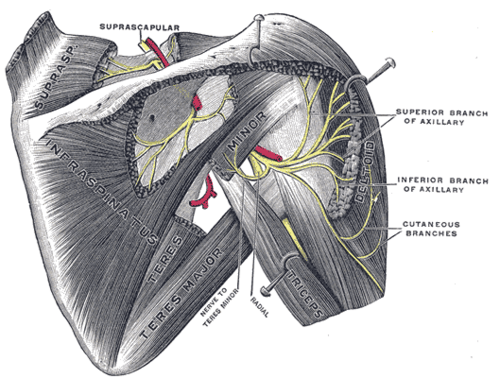
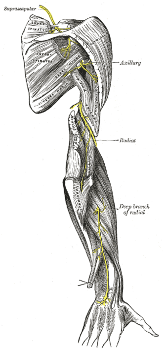
Branch of the posterior cord that passes through the quadrangular space around the surgical neck of the humerus to supply deltoid and teres minor.
Core anatomy
- Location
Posterior axilla → quadrangular space → deep surface of deltoid around the surgical neck of the humerus.
- Root values
C5, C6
- Origin
Terminal branch of the posterior cord of the brachial plexus.
- Course
Passes posteriorly through the quadrangular space with the posterior circumflex humeral artery. The space is bounded by teres minor above, teres major below, the long head of triceps brachii medially and the surgical neck of the humerus laterally. The nerve then winds around the surgical neck of the humerus, deep to deltoid.
- Branches
- Anterior branch: winds around the surgical neck deep to deltoid and supplies its anterior and middle parts
- Posterior branch: supplies teres minor and the posterior part of deltoid, then continues as the superior lateral cutaneous nerve of the arm
- Articular branch to the glenohumeral joint
- Motor
Deltoid and teres minor.
- Sensory
Skin over the inferior part of deltoid (the "regimental badge" area), via the superior lateral cutaneous nerve of the arm.
- Relations
The posterior circumflex humeral artery runs with it through the quadrangular space and around the surgical neck.
Muscles innervated
- Deltoid — axillary nerve (C5, C6)
- Teres minor — axillary nerve (C5, C6), via its posterior branch
Other structures innervated
Mentioned in
Generated automatically from links in other entries.


Clinical correlations
Fracture of the surgical neck of the humerus PANCE
- Mechanism / lesion
Usually a fall on an outstretched hand or directly onto the shoulder, commonly in older adults with osteoporosis. The nerve and the posterior circumflex humeral artery are in direct contact with the surgical neck.
- Classic presentation
Weak abduction (deltoid), especially from about 15° to 90°, and weak lateral rotation. Numbness over the regimental badge area. Deltoid atrophy develops later.
Anterior dislocation of the glenohumeral joint PANCE
- Mechanism / lesion
The dislocated humeral head, displaced anteroinferiorly, stretches or compresses the axillary nerve.
- Classic presentation
Same deficits as above. Check and document regimental badge sensation and deltoid function before and after reduction.
Quadrilateral space syndrome
- Mechanism / lesion
Compression of the axillary nerve (± posterior circumflex humeral artery) in the quadrangular space, e.g. by fibrous bands or muscle hypertrophy, typically in overhead athletes.
- Classic presentation
Poorly localized posterior shoulder pain and paresthesias, worse with abduction and external rotation. Teres minor ± deltoid atrophy may be seen on MRI.
High-yield PANCE pearls
- Surgical neck fracture or anterior dislocation → axillary nerve injury.
- Test regimental badge sensation (lateral shoulder) before and after reducing a shoulder dislocation.
- Quadrangular space contents: axillary nerve + posterior circumflex humeral artery.
- Deltoid atrophy → flattened shoulder contour; weak abduction beyond the first ~15°.
Imaging anatomy
- X-ray
The nerve is not visible. The shoulder trauma series (AP, scapular Y, axillary view) identifies surgical neck fractures and dislocations, the injuries that put the nerve at risk.
- MRI
Denervation of teres minor (± deltoid) appears as edema acutely and fatty atrophy chronically. Isolated teres minor atrophy suggests quadrilateral space syndrome. The neurovascular bundle can be followed through the quadrangular space on sagittal oblique images.
- Ultrasound
The nerve and posterior circumflex humeral artery can be identified together in the quadrangular space from a posterior approach, using color Doppler to find the artery.
- Key views & landmarks
- Shoulder trauma series: AP, scapular Y, axillary lateral
- MRI sagittal oblique through the quadrangular space
Physical exam
- Surface landmarks
The nerve lies deep to deltoid about at the level of the surgical neck, roughly 5–6 cm below the acromion. verify
- Strength / function testing
Resisted abduction with the arm at 90° (middle deltoid). Resisted lateral rotation with the elbow flexed at the side (teres minor, with infraspinatus). Sensation over the lateral shoulder (regimental badge).
Self-quiz
A 74-year-old woman falls onto her right shoulder. Radiographs show a fracture of the surgical neck of the humerus. Which additional finding is most likely?
Correct answer: B
The axillary nerve wraps around the surgical neck. Injury causes numbness over the inferior deltoid (the regimental badge area) and weak deltoid and teres minor. Wrist drop is the radial nerve at the midshaft (radial groove). Medial arm sensation is intercostobrachial/medial cutaneous nerve of the arm.
Show answer
B
The axillary nerve wraps around the surgical neck. Injury causes numbness over the inferior deltoid (the regimental badge area) and weak deltoid and teres minor. Wrist drop is the radial nerve at the midshaft (radial groove). Medial arm sensation is intercostobrachial/medial cutaneous nerve of the arm.
Which artery accompanies the axillary nerve through the quadrangular space?
Correct answer: E
The posterior circumflex humeral artery (from the third part of the axillary artery) travels with the axillary nerve. The circumflex scapular artery passes through the triangular space, and the profunda brachii runs with the radial nerve through the triangular interval.
Show answer
E
The posterior circumflex humeral artery (from the third part of the axillary artery) travels with the axillary nerve. The circumflex scapular artery passes through the triangular space, and the profunda brachii runs with the radial nerve through the triangular interval.
The lateral boundary of the quadrangular space is formed by which structure?
Correct answer: C
The quadrangular space is bounded by teres minor above, teres major below, the long head of triceps medially and the surgical neck of the humerus laterally.
Show answer
C
The quadrangular space is bounded by teres minor above, teres major below, the long head of triceps medially and the surgical neck of the humerus laterally.
A 22-year-old man is brought in with an anterior shoulder dislocation. Before reduction, which bedside test best screens for the nerve most commonly injured?
Correct answer: A
The axillary nerve is the nerve most commonly injured in anterior dislocation. Testing sensation over the regimental badge area is a quick screen, because deltoid strength is hard to test in a painful dislocated shoulder.
Show answer
A
The axillary nerve is the nerve most commonly injured in anterior dislocation. Testing sensation over the regimental badge area is a quick screen, because deltoid strength is hard to test in a painful dislocated shoulder.
A 25-year-old volleyball player has poorly localized posterior shoulder pain that worsens with abduction and external rotation. MRI shows isolated fatty atrophy of teres minor. Compression in which space is most likely?
Correct answer: E
Quadrilateral space syndrome compresses the axillary nerve (± posterior circumflex humeral artery) in the quadrangular space, typically in overhead athletes. Isolated teres minor atrophy is the classic MRI clue. Suprascapular and spinoglenoid notch lesions affect supraspinatus and/or infraspinatus instead.
Show answer
E
Quadrilateral space syndrome compresses the axillary nerve (± posterior circumflex humeral artery) in the quadrangular space, typically in overhead athletes. Isolated teres minor atrophy is the classic MRI clue. Suprascapular and spinoglenoid notch lesions affect supraspinatus and/or infraspinatus instead.
Flashcards
See also
- Glenohumeral joint
- Upper limb nerve injuries (comparison table)
- A shoulder forced back while blocking (clinical case)