Hip bone
Large flat bone formed by the fusion of the ilium, ischium and pubis at the acetabulum. Its palpable landmarks guide the exam, and its apophyses are classic sites of avulsion fractures in adolescents.
Core anatomy
- Location
The two hip bones form the lateral and anterior walls of the bony pelvis. Each articulates posteriorly with the sacrum and coccyx at the sacroiliac joint, anteriorly with its partner at the pubic symphysis, and laterally with the femoral head at the acetabulum.
- Classification
Irregular (flat) bone
- Key features & landmarks
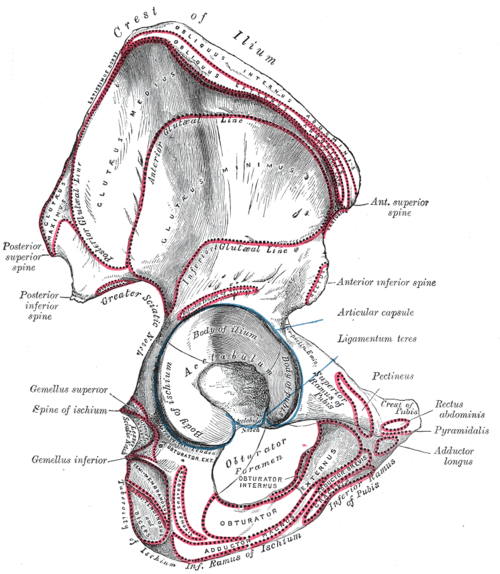
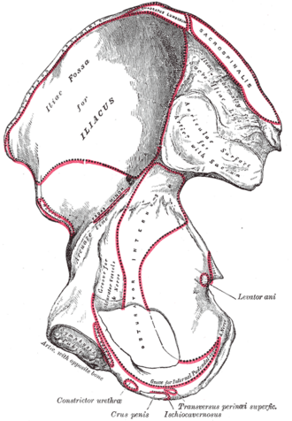
- Ilium: iliac crest (highest point at about L4), anterior superior iliac spine (ASIS; sartorius and the inguinal ligament), anterior inferior iliac spine (AIIS; straight head of rectus femoris and the iliofemoral ligament), posterior superior iliac spine (PSIS; at about S2), iliac fossa (iliacus), arcuate line, auricular surface for the sacrum
- Ischium: ischial spine (sacrospinous ligament; landmark for the pudendal nerve), lesser sciatic notch, ischial tuberosity (hamstrings and the sacrotuberous ligament; bears weight when sitting), ischial ramus
- Pubis: body, pubic tubercle (medial attachment of the inguinal ligament), pubic crest, superior ramus with the pectineal line, inferior ramus
- Greater sciatic notch: converted into the greater sciatic foramen by the sacrotuberous and sacrospinous ligaments
- Obturator foramen: closed by the obturator membrane except for the obturator canal (obturator nerve and vessels)
- Acetabulum: cup for the femoral head, formed by all three bones; the acetabular notch is bridged by the transverse acetabular ligament
- Ischiopubic ramus: joined inferior pubic and ischial rami; forms the side of the pubic arch and anchors the crura of the penis or clitoris
- Relations
The external iliac vessels and femoral nerve leave the pelvis over the superior pubic ramus under the inguinal ligament. The sciatic nerve exits through the greater sciatic notch below piriformis. The obturator nerve runs along the lateral pelvic wall to the obturator canal.
- Ossification
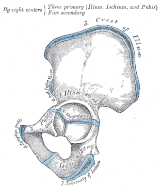
Three primary centers (ilium, ischium, pubis) appear before birth. They meet at the acetabulum in a Y-shaped triradiate cartilage that fuses around puberty (about 15–17 years) verify. Secondary centers include the iliac crest, the ASIS/AIIS, the ischial tuberosity and the pubic symphysis. The progress of iliac crest apophysis ossification (the Risser sign) is used to judge skeletal maturity in scoliosis.
Muscle attachments
- Adductor muscles of the thigh — origin
- Gluteus maximus — origin
- Gluteus medius and minimus — origin
- Hamstrings — origin
- Quadriceps femoris — origin
Joints
Mentioned in
Generated automatically from links in other entries.



Clinical correlations
Apophyseal avulsion fractures PANCE
- Mechanism / lesion
Forceful muscle contraction in adolescents (kicking, sprinting, jumping) pulls off an unfused apophysis: ASIS (sartorius), AIIS (rectus femoris), ischial tuberosity (hamstrings), iliac crest (abdominal muscles).
- Classic presentation
Sudden pain and a "pop" at the attachment, focal tenderness, and pain on resisted action of the muscle. Most are managed conservatively; widely displaced fragments (especially the ischial tuberosity) may need fixation.
Iliac crest contusion ("hip pointer")
- Mechanism / lesion
Direct blow to the iliac crest in contact sports.
- Classic presentation
Tenderness and bruising over the crest, pain with trunk rotation or side-bending. Managed conservatively.
Bone marrow aspiration and biopsy site
- Mechanism / lesion
The posterior iliac crest (near the PSIS) is the preferred site because it is superficial and away from major vessels.
- Classic presentation
Performed with the patient prone or in the lateral position.
Iliac crest bone graft harvest
- Mechanism / lesion
Graft is taken from the anterior or posterior crest.
- Classic presentation
Complications include donor-site pain and injury to the lateral femoral cutaneous nerve (numbness of the lateral thigh) when harvesting near the ASIS.
High-yield PANCE pearls
- Adolescent athlete + sudden hip/pelvic pain after kicking or sprinting → apophyseal avulsion (ASIS–sartorius, AIIS–rectus femoris, ischial tuberosity–hamstrings).
- Iliac crest = L4 (intercristal line); PSIS/dimples of Venus = S2.
- The ischial spine is the landmark for a pudendal nerve block.
- Posterior iliac crest is the usual site for bone marrow biopsy.
Imaging anatomy
- X-ray
AP pelvis shows the whole ring, both hip bones and the proximal femora. Judet (45° oblique) views show the anterior and posterior columns and walls of the acetabulum. Avulsions appear as a displaced crescent of bone next to the apophysis; compare with the other side.
- CT
CT with 3-D reconstruction is the standard for acetabular and complex pelvic fractures and for surgical planning.
- MRI
Detects occult fractures, stress fractures and apophysitis, and shows muscle–tendon injury at avulsion sites.
- Key views & landmarks
- AP pelvis
- Judet views (iliac and obturator obliques) for the acetabulum
- Inlet and outlet views for the pelvic ring
Physical exam
- Surface landmarks
Iliac crests (their tops are level with L4), ASIS at the anterior end of the crest, PSIS beneath the skin dimples above the buttocks (level of S2), pubic tubercle about 2–3 cm lateral to the midline on the pubic crest, and the ischial tuberosity deep in the lower buttock (easiest to feel with the hip flexed).
- Palpation
Palpate the crest from the ASIS to the PSIS for tenderness (hip pointer), and each apophysis in an adolescent with acute pain. Check that both ASIS are level (pelvic obliquity) with the patient standing.
- Strength / function testing
True leg length is measured from the ASIS to the medial malleolus; apparent leg length from the umbilicus to the medial malleolus.
Self-quiz
A 15-year-old soccer player feels a pop in the front of the hip while taking a hard shot on goal. There is tenderness just below the anterior superior iliac spine and pain on resisted hip flexion with the knee extended. Which muscle most likely avulsed its attachment?
Correct answer: B
The straight head of rectus femoris arises from the AIIS, just below the ASIS. Kicking loads rectus femoris strongly (hip flexion plus knee extension). Sartorius avulses the ASIS itself; iliopsoas avulses the lesser trochanter of the femur.
Show answer
B
The straight head of rectus femoris arises from the AIIS, just below the ASIS. Kicking loads rectus femoris strongly (hip flexion plus knee extension). Sartorius avulses the ASIS itself; iliopsoas avulses the lesser trochanter of the femur.
Which landmark is at the level of the second sacral vertebra?
Correct answer: C
The PSIS (marked by the skin dimples above the buttocks) lies at about S2, which is also roughly where the dural sac ends. The highest points of the iliac crests are at about L4.
Show answer
C
The PSIS (marked by the skin dimples above the buttocks) lies at about S2, which is also roughly where the dural sac ends. The highest points of the iliac crests are at about L4.
The three parts of the hip bone fuse at which structure?
Correct answer: C
The ilium, ischium and pubis meet at the acetabulum through the Y-shaped triradiate cartilage, which fuses around puberty.
Show answer
C
The ilium, ischium and pubis meet at the acetabulum through the Y-shaped triradiate cartilage, which fuses around puberty.
What is the preferred site for bone marrow aspiration and biopsy in adults?
Correct answer: C
The posterior iliac crest (near the PSIS) is preferred because it is superficial and away from major vessels. The procedure is done with the patient prone or lying on one side.
Show answer
C
The posterior iliac crest (near the PSIS) is preferred because it is superficial and away from major vessels. The procedure is done with the patient prone or lying on one side.
After an iliac crest bone graft is harvested near the anterior superior iliac spine, a patient has numbness over the lateral thigh. Which nerve was most likely injured?
Correct answer: D
The lateral femoral cutaneous nerve passes close to the ASIS, so harvesting graft from the anterior crest can injure it and cause numbness of the lateral thigh. Donor-site pain is the other common complication.
Show answer
D
The lateral femoral cutaneous nerve passes close to the ASIS, so harvesting graft from the anterior crest can injure it and cause numbness of the lateral thigh. Donor-site pain is the other common complication.