Bony pelvis
Ring formed by the two hip bones, the sacrum and the coccyx. Its inlet and outlet define the birth canal, and ring fractures from high-energy trauma can cause life-threatening hemorrhage.
Core anatomy
- Location
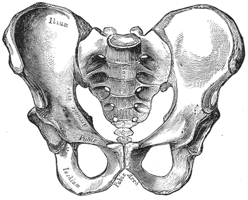
Between the lumbar spine and the lower limbs. Formed by the two hip bones (anteriorly and laterally) and the sacrum and coccyx and coccyx (posteriorly), joined at the sacroiliac joints and the pubic symphysis.
- Classification
Bony ring (pelvic girdle); strictly the pelvic girdle is the two hip bones and the sacrum.
- Key features & landmarks
- Pelvic inlet (brim): sacral promontory, ala of the sacrum, arcuate line of the ilium, pectineal line, pubic crest and the upper margin of the symphysis
- Greater (false) pelvis: above the brim, part of the abdominal cavity (iliac fossae)
- Lesser (true) pelvis: below the brim; contains the pelvic viscera and forms the bony birth canal
- Pelvic outlet: diamond-shaped, bounded by the pubic arch, ischial tuberosities, sacrotuberous ligaments and the tip of the coccyx
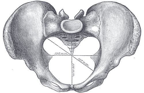
- Obstetric conjugate: sacral promontory to the nearest point on the back of the symphysis; the shortest AP diameter the fetal head must pass (about 10–11 cm)
- Diagonal conjugate: sacral promontory to the lower border of the symphysis; the one measured clinically (about 12.5 cm). The obstetric conjugate is about 1.5–2 cm shorter. verify
- Interspinous diameter: between the ischial spines (about 10 cm); usually the narrowest diameter of the midpelvis. The ischial spines also mark fetal station 0.
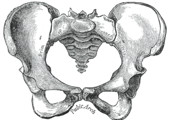
- Subpubic angle: wide in females (about 80–90° or more), narrow in males (about 50–60°)
- Relations
The lesser pelvis contains the urinary bladder, rectum and reproductive organs; its floor is the pelvic diaphragm. The lateral walls are lined by obturator internus; the posterior wall carries piriformis and the sacral plexus.
- Ossification
See hip bone and sacrum and coccyx.
- Notes
Female vs male pelvis: the female pelvis is lighter, with a round or oval inlet, a wider subpubic angle, a larger outlet, everted ischial tuberosities and less prominent ischial spines. The male inlet is heart-shaped. Caldwell–Moloy shapes are gynecoid (most favorable for vaginal delivery), android, anthropoid and platypelloid.
Mentioned in
Generated automatically from links in other entries.
Swipe sideways to see the whole diagram →



Clinical correlations
Pelvic ring fracture and hemorrhage PANCE
- Mechanism / lesion
High-energy trauma (motor vehicle collision, fall from height). A ring usually breaks in two places. Anteroposterior compression ("open book") widens the symphysis and disrupts the sacroiliac joints, enlarging pelvic volume. Lateral compression is the most common pattern. Vertical shear displaces a hemipelvis upward.
- Classic presentation
Pelvic pain, instability, perineal or scrotal bruising, and hemorrhagic shock. Bleeding is mostly venous (presacral and vesical plexuses) and from fracture surfaces; arterial bleeding (e.g. superior gluteal, internal pudendal, obturator from the internal iliac artery) is less common but may need embolization. Associated injuries include the urethra and bladder.
- Notes
Initial management: a pelvic binder centered over the greater trochanters, resuscitation, then angioembolization, preperitoneal packing or external fixation for ongoing bleeding. Do not repeatedly "spring" the pelvis to test stability, as this can dislodge clot.
Pubic ramus fracture in older adults PANCE
- Mechanism / lesion
A low-energy fall in an older adult with osteoporosis.
- Classic presentation
Groin pain and difficulty walking after a fall, often with normal hip X-rays at first. Usually treated with pain control and weight-bearing as tolerated. Look for an associated (often occult) sacral insufficiency fracture.
Cephalopelvic disproportion
- Mechanism / lesion
A fetal head too large for the maternal pelvis (or poorly positioned), including a contracted pelvis.
- Classic presentation
Arrest of labor despite adequate contractions; usually leads to cesarean delivery.
High-yield PANCE pearls
- Hypotensive trauma patient with a pelvic fracture → pelvic binder at the greater trochanters; bleeding is mostly venous.
- Pelvic fracture + blood at the urethral meatus → retrograde urethrogram before a Foley catheter.
- Obstetric conjugate = shortest AP diameter; diagonal conjugate is measured clinically.
- Ischial spines = fetal station 0 and the landmark for a pudendal block.
- Gynecoid pelvis is the most favorable for vaginal delivery.
Imaging anatomy
- X-ray
AP pelvis is a standard trauma adjunct in unstable patients. Inlet view (beam angled caudally) shows AP displacement; outlet view (angled cranially) shows vertical displacement and the sacral foramina. Symphyseal widening above about 2.5 cm suggests disruption of the pelvic floor and sacroiliac ligaments. verify
- CT
CT of the abdomen and pelvis with IV contrast is the definitive study in stable patients: fracture pattern, sacroiliac involvement, and an active contrast blush indicating arterial bleeding.
- MRI
Detects occult insufficiency fractures of the sacrum and pubic rami in older adults.
- Key views & landmarks
- AP pelvis (trauma series)
- Inlet and outlet views
- CT abdomen/pelvis with IV contrast
Physical exam
- Surface landmarks
Iliac crests, ASIS, pubic symphysis and tubercles, ischial tuberosities, PSIS and sacrum.
- Palpation
In trauma, gentle palpation of the pubic symphysis and iliac wings once only; avoid repeated compression. Inspect the perineum, scrotum and urethral meatus for blood, and perform a rectal (and in females, vaginal) exam for an open fracture.
- Strength / function testing
Clinical pelvimetry on bimanual exam estimates the diagonal conjugate, the prominence of the ischial spines and the subpubic angle.
Self-quiz
A 34-year-old man is brought in after a motorcycle collision. BP 78/50 mm Hg, pulse 136/min. The pelvis is unstable to gentle compression and a FAST exam is negative. What is the most appropriate immediate step?
Correct answer: A
Hemodynamic instability with an unstable pelvis and negative FAST suggests pelvic hemorrhage. A binder over the greater trochanters reduces pelvic volume and tamponades venous bleeding while resuscitation continues. A Foley should wait until urethral injury is excluded, and repeated compression can dislodge clot.
Show answer
A
Hemodynamic instability with an unstable pelvis and negative FAST suggests pelvic hemorrhage. A binder over the greater trochanters reduces pelvic volume and tamponades venous bleeding while resuscitation continues. A Foley should wait until urethral injury is excluded, and repeated compression can dislodge clot.
Which pelvic diameter is the shortest anteroposterior diameter through which the fetal head must pass?
Correct answer: B
The obstetric conjugate runs from the sacral promontory to the closest point on the posterior surface of the symphysis. It can't be measured directly, so it is estimated by subtracting about 1.5–2 cm from the diagonal conjugate measured on exam. The interspinous diameter is transverse, not AP.
Show answer
B
The obstetric conjugate runs from the sacral promontory to the closest point on the posterior surface of the symphysis. It can't be measured directly, so it is estimated by subtracting about 1.5–2 cm from the diagonal conjugate measured on exam. The interspinous diameter is transverse, not AP.
Compared with the male pelvis, the female pelvis typically has which feature?
Correct answer: C
The female pelvis is adapted for childbirth: a wider subpubic angle, an oval inlet, a larger outlet, and less prominent ischial spines. A heart-shaped inlet and narrow subpubic angle are male (android) features.
Show answer
C
The female pelvis is adapted for childbirth: a wider subpubic angle, an oval inlet, a larger outlet, and less prominent ischial spines. A heart-shaped inlet and narrow subpubic angle are male (android) features.
In a hemodynamically unstable patient with a pelvic ring fracture, where does most of the bleeding come from?
Correct answer: B
Pelvic fracture bleeding is mostly venous (presacral and vesical plexuses) and from the fracture surfaces. Arterial bleeding from internal iliac branches (superior gluteal, internal pudendal, obturator) is less common but may need angioembolization. A pelvic binder over the greater trochanters reduces pelvic volume.
Show answer
B
Pelvic fracture bleeding is mostly venous (presacral and vesical plexuses) and from the fracture surfaces. Arterial bleeding from internal iliac branches (superior gluteal, internal pudendal, obturator) is less common but may need angioembolization. A pelvic binder over the greater trochanters reduces pelvic volume.
During labor, fetal station 0 is defined by the level of which bony landmark?
Correct answer: A
The ischial spines mark fetal station 0. The distance between them (the interspinous diameter) is usually the narrowest diameter of the midpelvis, and they are also the landmark for a pudendal nerve block.
Show answer
A
The ischial spines mark fetal station 0. The distance between them (the interspinous diameter) is usually the narrowest diameter of the midpelvis, and they are also the landmark for a pudendal nerve block.
Flashcards
See also
- Hip bone
- Sacrum and coccyx
- Sacroiliac joint
- Pubic symphysis
- An older woman who can't stand after a fall (clinical case)
- Blood at the urethral meatus (clinical case)