Mandible
The lower jaw — the largest facial bone and the only movable bone of the skull. As a ring with the skull base, it often fractures in two places, and the inferior alveolar nerve runs through it.
Core anatomy
- Location
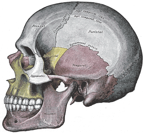
Lower face, articulating with the temporal bones at the temporomandibular joints.
- Classification
Irregular bone (body and two rami)
- Key features & landmarks
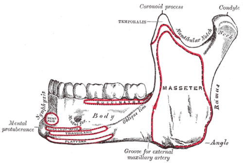
- Body: alveolar process holding the lower teeth; mental foramen (below the 2nd premolar) transmitting the mental nerve and vessels; symphysis in the midline
- Ramus: vertical plate with the angle at its lower back corner; the mandibular foramen on its inner surface (inferior alveolar nerve and vessels), guarded by the lingula
- Condylar process: head (condyle) and neck; articulates with the mandibular fossa of the temporal bone
- Coronoid process: insertion of temporalis
- Attachments: masseter and medial pterygoid at the angle; lateral pterygoid at the neck of the condyle; digastric, geniohyoid and genioglossus near the symphysis
- Relations
The inferior alveolar nerve (from V3) runs in the mandibular canal to the teeth and exits as the mental nerve to the lower lip and chin.
- Ossification
Mostly intramembranous (around Meckel cartilage of the first pharyngeal arch); the two halves fuse at the symphysis in the first year.
Joints
Forms a boundary of
Mentioned in
Generated automatically from links in other entries.


Clinical correlations
Mandibular fractures PANCE
- Mechanism / lesion
Blunt trauma (assault, falls, motor vehicle collisions). Because the mandible and skull base form a ring, fractures are often bilateral (e.g. body or angle on one side and condyle on the other). Most common sites include the condyle, angle and body.
- Classic presentation
Malocclusion, pain and trismus, step deformity, sublingual hematoma (highly suggestive), and numbness of the lower lip and chin (inferior alveolar/mental nerve injury). Fractures through tooth sockets are open fractures → antibiotics.
Inferior alveolar nerve block
- Mechanism / lesion
Local anesthetic near the mandibular foramen on the inner ramus.
- Classic presentation
Anesthesia of the lower teeth, lower lip and chin on that side (the lingual nerve is often blocked too — numb tongue).
Medication-related osteonecrosis of the jaw
- Mechanism / lesion
Associated with antiresorptive drugs (bisphosphonates, denosumab), especially IV/high-dose, and dental extractions.
- Classic presentation
Exposed necrotic bone in the mouth for over 8 weeks; mandible more than maxilla.
High-yield PANCE pearls
- Mandible fractures are often bilateral (ring) — look for a second fracture.
- Sublingual hematoma after facial trauma → mandibular fracture.
- Lower lip/chin numbness = inferior alveolar/mental nerve (V3).
- Bisphosphonates + dental extraction → osteonecrosis of the jaw.
Imaging anatomy
- X-ray
A panoramic radiograph (Panorex) shows the whole mandible; supplemented by PA mandible views.
- CT
CT of the face (maxillofacial CT) is the standard for mandibular and midface fractures and surgical planning.
- Key views & landmarks
- Panorex
- Maxillofacial CT with 3-D reconstruction
Physical exam
- Surface landmarks
Angle of the mandible, mental protuberance, condyle palpable in front of the tragus.
- Palpation
Palpate along the inferior border for steps; check occlusion ("does your bite feel normal?"), mouth opening and sensation of the lower lip and chin.
Tongue blade test
- Technique
The patient bites down on a tongue blade while the examiner twists it.
- Positive test
The patient can't hold the blade and it can't be broken (inability to grip).
- Significance
Suggests mandibular fracture; being able to break the blade makes a fracture less likely.
Self-quiz
After a punch to the left jaw, a patient has malocclusion, a step in the left mandibular body and pain in front of the right ear. What is the most likely explanation for the right-sided pain?
Correct answer: B
The mandible and skull base form a ring, so fractures are often bilateral — a body fracture on the struck side with a contralateral condylar fracture is a classic combination.
Show answer
B
The mandible and skull base form a ring, so fractures are often bilateral — a body fracture on the struck side with a contralateral condylar fracture is a classic combination.
Which nerve passes through the mental foramen?
Correct answer: B
The inferior alveolar nerve (V3) runs through the mandibular canal and exits at the mental foramen as the mental nerve, supplying the skin of the lower lip and chin.
Show answer
B
The inferior alveolar nerve (V3) runs through the mandibular canal and exits at the mental foramen as the mental nerve, supplying the skin of the lower lip and chin.
After a fist fight, a man has malocclusion and a bruise under his tongue. A tooth socket lies in the fracture line. Besides fixation, what does this need?
Correct answer: B
A sublingual hematoma after facial trauma strongly suggests a mandibular fracture, and a fracture through a tooth socket communicates with the mouth, so it is an open fracture that needs antibiotics.
Show answer
B
A sublingual hematoma after facial trauma strongly suggests a mandibular fracture, and a fracture through a tooth socket communicates with the mouth, so it is an open fracture that needs antibiotics.