Kidney
Paired retroperitoneal organs at T12–L3, with the right kidney slightly lower than the left. Stones, pyelonephritis, renal cell carcinoma and trauma are the key clinical problems.
Core anatomy
- Location
Retroperitoneal, on the posterior abdominal wall beside the vertebral column from about T12 to L3. The right kidney lies slightly lower (displaced by the liver). The hila lie at about L1 (transpyloric plane). The kidneys move a few centimeters with respiration.
- Parts
- Cortex, medulla (renal pyramids), renal columns; minor and major calyces → renal pelvis → ureter
- Hilum, anterior to posterior: renal vein, renal artery, renal pelvis (VAP)
- Coverings, inside to out: fibrous capsule, perirenal fat, renal (Gerota) fascia (also encloses the suprarenal gland), pararenal fat
- Relations
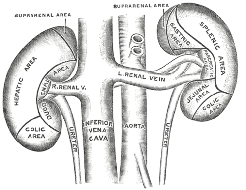
Posterior (both): diaphragm, 12th rib (and 11th on the left), psoas major, quadratus lumborum, transversus abdominis; subcostal, iliohypogastric and ilioinguinal nerves. Anterior, right: liver, 2nd part of the duodenum, hepatic flexure. Anterior, left: stomach, spleen, tail of the pancreas, splenic flexure, jejunum. Superiorly, the suprarenal gland.
- Arterial supply
Renal arteries from the abdominal aorta at about L1–L2 (the right is longer, passing behind the IVC). Segmental arteries are end arteries (so occlusion causes segmental infarction). Accessory renal arteries are common.
- Venous drainage
Renal veins → inferior vena cava. The left renal vein is longer, crosses anterior to the aorta beneath the SMA, and receives the left gonadal and left suprarenal veins.
- Lymphatic drainage
Lumbar (para-aortic) nodes.
- Innervation
Renal plexus (sympathetic from the lesser/least splanchnic nerves, about T10–L1). Pain from capsular or pelvic distension refers to the flank ("loin") and groin.

Clinical correlations
Nephrolithiasis (kidney stones) PANCE
- Mechanism / lesion
Calcium oxalate stones are most common; also calcium phosphate, uric acid (radiolucent), struvite (urease-producing organisms, staghorn), and cystine.
- Classic presentation
Sudden severe colicky flank pain radiating to the groin, restlessness, nausea, hematuria. Stones lodge at the ureter's narrow points (see ureter). Infection above an obstructing stone is a urologic emergency (decompression needed).
Pyelonephritis PANCE
- Mechanism / lesion
Ascending bacterial infection (most often E. coli) from the bladder.
- Classic presentation
Fever, flank pain, costovertebral angle tenderness, nausea, with or without cystitis symptoms. Urinalysis shows pyuria and WBC casts.
Renal cell carcinoma PANCE
- Mechanism / lesion
Most common renal malignancy in adults. Risk factors include smoking, obesity, hypertension and von Hippel–Lindau disease.
- Classic presentation
Often incidental on imaging. Classic (uncommon) triad: hematuria, flank pain, palpable mass. Paraneoplastic syndromes: polycythemia (EPO), hypercalcemia, hypertension. Invasion of the left renal vein can cause a left varicocele; tumor thrombus may extend into the IVC.
Horseshoe kidney
- Mechanism / lesion
Fusion of the lower poles; ascent is arrested by the inferior mesenteric artery. Associated with Turner syndrome.
- Classic presentation
Usually asymptomatic; increased risk of obstruction, stones, infection and trauma.
High-yield PANCE pearls
- Hilum order anterior → posterior: vein, artery, pelvis (VAP).
- Stone workup: non-contrast CT (ultrasound in pregnancy and young patients); uric acid stones are radiolucent.
- Pyelonephritis: fever + CVA tenderness + WBC casts.
- RCC: hematuria + flank pain + mass; polycythemia; left varicocele.
- Right kidney sits lower than the left (liver).
Imaging anatomy
- X-ray
KUB shows radiopaque stones (calcium, struvite, cystine); uric acid stones are radiolucent. Staghorn calculi fill the collecting system.
- CT
Non-contrast CT is the most sensitive test for stones (size, location, hydronephrosis). CT urography (multiphase with contrast) evaluates hematuria and masses. Renal masses are characterized by enhancement on multiphase CT or MRI.
- MRI
Characterizes indeterminate renal masses and renal vein/IVC tumor thrombus; avoids iodinated contrast.
- Ultrasound
First-line for hydronephrosis, renal size, cysts vs solid masses, and stones in pregnancy or children. Normal adult kidney length is roughly 10–12 cm. verify Doppler for renal artery stenosis and transplant perfusion.
- Key views & landmarks
- Non-contrast CT abdomen/pelvis (stone protocol)
- Renal ultrasound
- CT urogram for hematuria
Physical exam
- Surface landmarks
The kidneys lie under the 11th–12th ribs posteriorly. The costovertebral angle (between the 12th rib and the vertebral column) overlies the kidney.
- Palpation
Bimanual palpation (ballottement): one hand posteriorly under the flank, the other anteriorly below the costal margin; push up from behind during deep inspiration. The lower pole of the right kidney may be palpable in thin people.
- Strength / function testing
CVA tenderness: place one hand over the costovertebral angle and strike it with the ulnar side of the other fist. Pain suggests pyelonephritis or obstruction. Auscultate for a renal artery bruit (hypertension workup).
Self-quiz
A 34-year-old man has sudden severe left flank pain radiating to the groin, is writhing, and has microscopic hematuria. What is the most sensitive imaging test?
Correct answer: B
Non-contrast CT is the most sensitive test for urinary stones and shows hydronephrosis. Ultrasound is preferred in pregnancy.
Show answer
B
Non-contrast CT is the most sensitive test for urinary stones and shows hydronephrosis. Ultrasound is preferred in pregnancy.
At the renal hilum, what is the arrangement of structures from anterior to posterior?
Correct answer: B
From anterior to posterior: renal vein, renal artery, renal pelvis (VAP).
Show answer
B
From anterior to posterior: renal vein, renal artery, renal pelvis (VAP).
A 60-year-old smoker has hematuria, an elevated hematocrit and a new left-sided varicocele. What is the most likely diagnosis?
Correct answer: B
Renal cell carcinoma can secrete EPO (polycythemia) and invade the left renal vein, which receives the left gonadal vein, causing a left varicocele.
Show answer
B
Renal cell carcinoma can secrete EPO (polycythemia) and invade the left renal vein, which receives the left gonadal vein, causing a left varicocele.
Which finding on urinalysis best distinguishes pyelonephritis from cystitis?
Correct answer: C
WBC casts form in the renal tubules and indicate upper tract (kidney) involvement such as pyelonephritis.
Show answer
C
WBC casts form in the renal tubules and indicate upper tract (kidney) involvement such as pyelonephritis.
Which kidney usually lies lower, and why?
Correct answer: D
The right kidney sits slightly lower because the liver displaces it. The kidneys lie from about T12 to L3, with the hila at about L1 (transpyloric plane).
Show answer
D
The right kidney sits slightly lower because the liver displaces it. The kidneys lie from about T12 to L3, with the hila at about L1 (transpyloric plane).