Paranasal sinuses
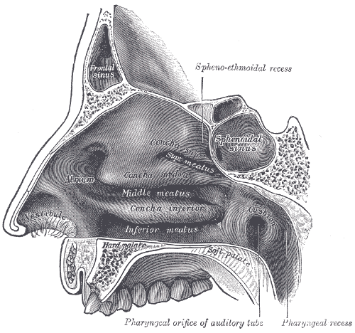
Four paired air-filled cavities — frontal, ethmoid, sphenoid and maxillary — draining into the nasal cavity. Acute sinusitis is usually viral; ethmoid and frontal infections can spread to the orbit and brain.
Core anatomy
- Location
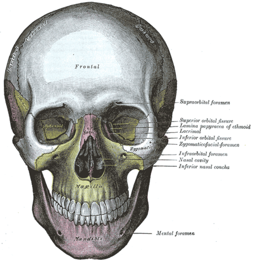
Within the frontal, ethmoid, sphenoid and maxillary bones around the nasal cavity.
- Boundaries
- frontal sinus
- In the frontal bone above the orbits; the anterior cranial fossa lies behind its posterior wall
- ethmoid cells
- Between the orbits (lamina papyracea laterally — paper-thin) and the nasal cavity
- sphenoid sinus
- In the sphenoid body below the pituitary (sella turcica); cavernous sinuses and internal carotid arteries lie laterally
- maxillary sinus
- The largest; in the maxilla below the orbit (orbital floor above), roots of the upper molars below
- Contents
- Air and a thin mucoperiosteal lining with cilia that sweep mucus toward the ostia
- Communications
Middle meatus (ostiomeatal complex): frontal, maxillary and anterior ethmoid. Superior meatus: posterior ethmoid. Sphenoethmoidal recess: sphenoid. The maxillary ostium is high on its medial wall, so drainage depends on cilia rather than gravity.
- Notes
At birth only the maxillary and ethmoid sinuses are present; the sphenoid develops in childhood and the frontal sinus appears around age 5–7 and develops through adolescence. verify


Clinical correlations
Acute rhinosinusitis PANCE
- Mechanism / lesion
Usually viral (URI); bacterial (S. pneumoniae, H. influenzae, M. catarrhalis) in a minority.
- Classic presentation
Nasal congestion, purulent discharge, facial pain/pressure (worse bending forward), maxillary toothache. Suspect bacterial if symptoms last ≥10 days without improvement, are severe (fever ≥39 °C with purulent discharge for 3–4 days), or "double-worsen" after initial improvement → amoxicillin-clavulanate. Otherwise symptomatic care (saline irrigation, intranasal steroids).
Complications of sinusitis PANCE
- Mechanism / lesion
Spread through thin bone and valveless veins.
- Classic presentation
Orbital cellulitis (ethmoid — see orbit), Pott puffy tumor (frontal bone osteomyelitis with forehead swelling), meningitis, epidural/brain abscess, cavernous sinus thrombosis (sphenoid/ethmoid — ophthalmoplegia, proptosis, fever).
Chronic rhinosinusitis
- Mechanism / lesion
Inflammation lasting over 12 weeks, with or without nasal polyps.
- Classic presentation
Congestion, discharge, facial pressure, reduced smell; confirmed with nasal endoscopy or CT; intranasal steroids and saline irrigation, surgery if refractory.
High-yield PANCE pearls
- Bacterial sinusitis if ≥10 days, severe, or double-worsening → amoxicillin-clavulanate.
- Most acute sinusitis is viral — no antibiotics, no imaging.
- Ethmoid sinusitis → orbital cellulitis (children).
- Frontal sinusitis + forehead swelling → Pott puffy tumor.
- Maxillary sinusitis can cause upper toothache (and vice versa).
Imaging anatomy
- X-ray
Sinus X-rays are not recommended.
- CT
CT of the sinuses without contrast for chronic or recurrent sinusitis or before surgery; contrast CT (± MRI) when complications are suspected. Not for uncomplicated acute sinusitis.
- MRI
Intracranial and orbital complications, fungal sinusitis, tumors.
- Key views & landmarks
- Non-contrast CT sinuses (coronal)
Physical exam
- Surface landmarks
Frontal sinus above the medial eyebrow; maxillary sinus over the cheek below the orbit.
- Palpation
Percussion or palpation tenderness over the sinuses (limited accuracy).
- Strength / function testing
Anterior rhinoscopy for purulent discharge in the middle meatus; check eye movements and vision if orbital spread is possible; transillumination is rarely used.
Self-quiz
A 34-year-old has had nasal congestion, purulent discharge and maxillary pain for 5 days. She is afebrile and improving slightly. What is the most appropriate management?
Correct answer: C
At 5 days and improving, this is most likely viral rhinosinusitis. Antibiotics are for symptoms lasting 10 days or more, severe illness, or double-worsening. Imaging is not needed for uncomplicated sinusitis.
Show answer
C
At 5 days and improving, this is most likely viral rhinosinusitis. Antibiotics are for symptoms lasting 10 days or more, severe illness, or double-worsening. Imaging is not needed for uncomplicated sinusitis.
Which sinus is most often the source of orbital cellulitis in children?
Correct answer: C
The ethmoid cells are separated from the orbit only by the paper-thin lamina papyracea, so ethmoid sinusitis is the usual source of orbital cellulitis.
Show answer
C
The ethmoid cells are separated from the orbit only by the paper-thin lamina papyracea, so ethmoid sinusitis is the usual source of orbital cellulitis.
Which feature suggests bacterial rather than viral acute rhinosinusitis?
Correct answer: B
Suspect bacterial sinusitis if symptoms last ≥ 10 days without improvement, are severe (fever ≥ 39 °C with purulent discharge for 3–4 days) or "double-worsen"; treat with amoxicillin-clavulanate. Most acute sinusitis is viral.
Show answer
B
Suspect bacterial sinusitis if symptoms last ≥ 10 days without improvement, are severe (fever ≥ 39 °C with purulent discharge for 3–4 days) or "double-worsen"; treat with amoxicillin-clavulanate. Most acute sinusitis is viral.
A teenager with frontal sinusitis develops a soft, tender swelling of the forehead. What is the most likely complication?
Correct answer: E
Pott puffy tumor is frontal bone osteomyelitis with a subperiosteal abscess, producing forehead swelling; it can extend intracranially and needs CT and IV antibiotics.
Show answer
E
Pott puffy tumor is frontal bone osteomyelitis with a subperiosteal abscess, producing forehead swelling; it can extend intracranially and needs CT and IV antibiotics.
Why does the maxillary sinus drain poorly and depend on ciliary action?
Correct answer: A
The maxillary ostium is high on the medial wall, opening into the middle meatus, so drainage depends on cilia rather than gravity. Maxillary sinusitis can also cause upper toothache.
Show answer
A
The maxillary ostium is high on the medial wall, opening into the middle meatus, so drainage depends on cilia rather than gravity. Maxillary sinusitis can also cause upper toothache.