Piriformis
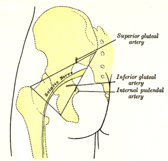
Pear-shaped lateral rotator of the hip and the key landmark of the gluteal region — structures leave the pelvis above or below it. It can irritate the sciatic nerve (piriformis syndrome).
Core anatomy
- Location
Leaves the pelvis through the greater sciatic foramen, which it almost fills, deep to gluteus maximus.
- Group
Gluteal region, deep layer (short lateral rotators)
- Origin
Anterior (pelvic) surface of the sacrum and coccyx (S2–S4 region) and the sacrotuberous ligament.
- Insertion
Upper border of the greater trochanter of the femur.
- Innervation
Nerve to piriformis (S1–S2), from the sacral plexus
- Blood supply
Superior and inferior gluteal arteries and the lateral sacral arteries.
- Action
- Lateral rotation of the extended hip
- Abduction of the flexed hip
- Holds the femoral head in the acetabulum
- Relations
Above piriformis: superior gluteal nerve and vessels. Below piriformis: Sciatic nerve, inferior gluteal nerve and vessels, posterior femoral cutaneous nerve, pudendal nerve and internal pudendal vessels, and the nerves to obturator internus and quadratus femoris. The sciatic nerve (or its common fibular division) passes through or above the muscle in a minority of people.

Clinical correlations
Piriformis syndrome
- Mechanism / lesion
Irritation or compression of the sciatic nerve by piriformis (spasm, hypertrophy, anatomic variants, trauma); a diagnosis of exclusion.
- Classic presentation
Deep buttock pain radiating down the back of the thigh, worse with prolonged sitting and hip flexion–adduction– internal rotation; tenderness over the muscle; no back pain or root signs. Treat with stretching and physical therapy; injection if refractory. verify
High-yield PANCE pearls
- Piriformis is the landmark of the gluteal region: superior gluteal vessels/nerve above, everything else below.
- Buttock pain worse with sitting, normal back exam → consider piriformis syndrome (exclude radiculopathy first).
Imaging anatomy
- MRI
Excludes lumbar disc herniation and shows piriformis asymmetry, variants or masses; MR neurography for the sciatic nerve.
- Ultrasound
Guides injections.
Physical exam
- Surface landmarks
Lies along a line from the upper sacrum (about 2 cm lateral to the midline at S2) to the top of the greater trochanter.
- Palpation
Deep tenderness in the buttock along that line.
FAIR test
- Technique
Side-lying with the tested side up; the hip is flexed, adducted and internally rotated.
- Positive test
Buttock pain or sciatic symptoms.
- Significance
Suggests piriformis syndrome (limited specificity).
Self-quiz
Which structure passes through the greater sciatic foramen above the piriformis muscle?
Correct answer: C
Only the superior gluteal nerve and vessels pass above piriformis; all other nerves and vessels leave the pelvis below it.
Show answer
C
Only the superior gluteal nerve and vessels pass above piriformis; all other nerves and vessels leave the pelvis below it.
What is the main action of piriformis with the hip extended?
Correct answer: B
Piriformis is one of the short lateral rotators of the hip (with the obturators, gemelli and quadratus femoris). With the hip flexed beyond about 60–90°, it acts as an abductor (and its rotation reverses).
Show answer
B
Piriformis is one of the short lateral rotators of the hip (with the obturators, gemelli and quadratus femoris). With the hip flexed beyond about 60–90°, it acts as an abductor (and its rotation reverses).
Which structure leaves the pelvis through the greater sciatic foramen below piriformis in most people?
Correct answer: D
Piriformis is the landmark of the gluteal region: only the superior gluteal nerve and vessels pass above it. The sciatic nerve, inferior gluteal nerve and vessels, posterior femoral cutaneous nerve and pudendal nerve pass below it.
Show answer
D
Piriformis is the landmark of the gluteal region: only the superior gluteal nerve and vessels pass above it. The sciatic nerve, inferior gluteal nerve and vessels, posterior femoral cutaneous nerve and pudendal nerve pass below it.