Circumflex artery
Branch of the left coronary artery in the left atrioventricular groove that supplies the lateral wall of the left ventricle and the left atrium; its occlusion causes lateral (and sometimes posterior) MI.
Core anatomy
- Location
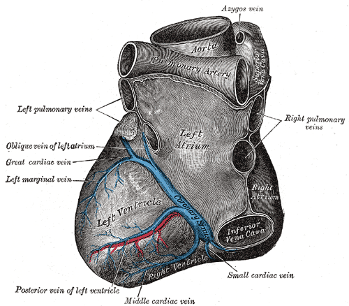
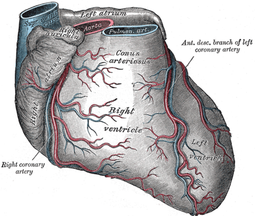
Left atrioventricular (coronary) groove, curving around the left (obtuse) margin to the posterior surface.
- Origin
- Course
Runs in the left AV groove around the obtuse margin toward the posterior heart. In left-dominant hearts it continues to give the posterior interventricular artery.
- Branches
- Left (obtuse) marginal arteries: to the lateral LV wall
- Atrial branches; the SA nodal artery in about 40%
- Posterior interventricular artery in left-dominant hearts
- Supplies
Left atrium, the lateral (and posterolateral) wall of the LV, the anterolateral papillary muscle (shared with the LAD), and in left dominance the posterior septum and inferior wall (including the AV node).
- Relations
Runs alongside the great cardiac vein/coronary sinus in the AV groove, close to the mitral annulus.
Structures supplied
Mentioned in
Generated automatically from links in other entries.


Clinical correlations
Lateral myocardial infarction PANCE
- Mechanism / lesion
Occlusion of the circumflex or an obtuse marginal branch.
- Classic presentation
ST elevation in I, aVL, V5–V6. Circumflex occlusions are often electrically silent on the standard 12-lead ECG because the lateral/posterior wall is poorly represented.
Posterior myocardial infarction PANCE
- Mechanism / lesion
Occlusion of the circumflex (or of a dominant RCA's posterolateral branches), often with inferior or lateral MI.
- Classic presentation
ST depression with tall R waves in V1–V3 (the mirror image of posterior ST elevation). Confirm with posterior leads V7–V9 showing ST elevation.
High-yield PANCE pearls
- Lateral MI: I, aVL, V5–V6 → circumflex.
- ST depression + tall R in V1–V3 → think posterior MI; get V7–V9.
- Circumflex occlusion can be ECG-silent; rely on symptoms, troponin and echo.
Imaging anatomy
- CT
Coronary CTA; the circumflex runs near the mitral annulus.
- Ultrasound
Echo shows lateral and posterolateral wall motion abnormalities; may show MR from papillary muscle dysfunction.
- Key views & landmarks
- Invasive angiography (RAO caudal view shows the circumflex and obtuse marginals)
- Posterior leads V7–V9
Physical exam
- Strength / function testing
Listen for a new MR murmur (papillary muscle dysfunction) after lateral or posterior MI.
Self-quiz
A patient has ST elevation in I, aVL, V5 and V6. Which artery is most likely occluded?
Correct answer: C
I, aVL, V5–V6 are the lateral leads, the circumflex territory (or a large diagonal of the LAD).
Show answer
C
I, aVL, V5–V6 are the lateral leads, the circumflex territory (or a large diagonal of the LAD).
A patient with chest pain has horizontal ST depression and tall R waves in V1–V3. What is the next best diagnostic step?
Correct answer: B
ST depression with tall R waves in V1–V3 may be a posterior STEMI seen from the front. Leads V7–V9 show the ST elevation directly.
Show answer
B
ST depression with tall R waves in V1–V3 may be a posterior STEMI seen from the front. Leads V7–V9 show the ST elevation directly.
In a left-dominant heart, which artery gives rise to the posterior interventricular artery?
Correct answer: B
In left dominance, the circumflex continues around the posterior AV groove to give the posterior interventricular artery.
Show answer
B
In left dominance, the circumflex continues around the posterior AV groove to give the posterior interventricular artery.
In which groove does the circumflex artery run?
Correct answer: A
The circumflex runs in the left AV groove around the obtuse margin, alongside the great cardiac vein and coronary sinus and close to the mitral annulus. The LAD lies in the anterior interventricular groove and the RCA in the right AV groove.
Show answer
A
The circumflex runs in the left AV groove around the obtuse margin, alongside the great cardiac vein and coronary sinus and close to the mitral annulus. The LAD lies in the anterior interventricular groove and the RCA in the right AV groove.
Why can a circumflex occlusion be missed on a standard 12-lead ECG?
Correct answer: D
The circumflex supplies the lateral and posterolateral LV wall, which the standard 12 leads represent poorly, so occlusion can be electrically silent. Rely on symptoms, troponin, echocardiography, and posterior leads (V7–V9).
Show answer
D
The circumflex supplies the lateral and posterolateral LV wall, which the standard 12 leads represent poorly, so occlusion can be electrically silent. Rely on symptoms, troponin, echocardiography, and posterior leads (V7–V9).