Inferior epigastric artery
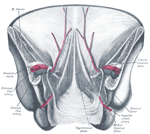
Branch of the external iliac artery that ascends into the rectus sheath. It's the landmark separating indirect from direct inguinal hernias and must be avoided during paracentesis and laparoscopic port placement.
Core anatomy
- Location
Deep surface of the lower anterior abdominal wall, running from just above the inguinal ligament into the rectus sheath.
- Origin
External iliac artery, just superior to the inguinal ligament.
- Course
Runs superomedially in the extraperitoneal tissue, medial to the deep inguinal ring, raising a peritoneal fold (the lateral umbilical fold). It pierces the transversalis fascia and enters the rectus sheath near the arcuate line, ascending on the deep surface of rectus abdominis.
- Branches
- Cremasteric artery (to the spermatic cord) or artery of the round ligament
- Pubic branch (may anastomose with the obturator artery, the "corona mortis")
- Muscular and cutaneous branches
- Supplies
Lower rectus abdominis and the medial anterior abdominal wall.
- Anastomoses
With the superior epigastric artery (from the internal thoracic) inside the rectus sheath, a collateral route between the subclavian and iliac systems (important in coarctation and aortoiliac occlusion).
- Relations
Forms the lateral border of the inguinal (Hesselbach) triangle (inguinal triangle). Indirect inguinal hernias emerge lateral to it through the deep ring; direct hernias emerge medial to it.
Structures supplied
Found in
Forms a boundary of
Generated automatically from links in other entries.
Swipe sideways to see the whole diagram →

Clinical correlations
Landmark for inguinal hernia type PANCE
- Mechanism / lesion
The vessels mark the medial edge of the deep inguinal ring.
- Classic presentation
At surgery (especially laparoscopic), a hernia sac lateral to the vessels is indirect and one medial to them is direct.
Injury during paracentesis or laparoscopy PANCE
- Mechanism / lesion
Needles or trocars placed too medially in the lower abdomen.
- Classic presentation
Abdominal wall hematoma or hemoperitoneum. Paracentesis is performed lateral to the rectus sheath, commonly in the left lower quadrant about two fingerbreadths medial and superior to the ASIS, ideally with ultrasound guidance to avoid the vessels. verify
Rectus sheath hematoma
- Mechanism / lesion
Rupture of the inferior epigastric artery, often in anticoagulated patients.
- Classic presentation
Tender lower abdominal wall mass (see rectus abdominis).
High-yield PANCE pearls
- Indirect hernia = lateral to the inferior epigastric vessels; direct = medial (Hesselbach triangle).
- Paracentesis: stay lateral to the rectus sheath (LLQ preferred) to avoid the inferior epigastric artery.
- Superior–inferior epigastric anastomosis = collateral pathway in coarctation or aortoiliac occlusion.
Imaging anatomy
- CT
Contrast CT shows the vessel and active bleeding in rectus sheath hematoma; CT angiography before abdominal wall flaps (the DIEP flap in breast reconstruction is based on its perforators).
- Ultrasound
Color Doppler identifies the vessel before paracentesis or trocar placement.
- Key views & landmarks
- Color Doppler of the lower abdominal wall before procedures
Physical exam
- Surface landmarks
Runs roughly from the mid-inguinal point toward the lateral edge of the rectus sheath at the arcuate line. Not palpable.
- Strength / function testing
Before paracentesis, percuss for dullness laterally and use ultrasound to find fluid and avoid the vessels.
Self-quiz
During laparoscopic hernia repair, a hernia sac is found passing through the abdominal wall medial to the inferior epigastric vessels. What type of hernia is it?
Correct answer: B
Direct hernias pass through the inguinal (Hesselbach) triangle, medial to the inferior epigastric vessels. Indirect hernias enter the deep ring lateral to them; femoral hernias pass below the inguinal ligament.
Show answer
B
Direct hernias pass through the inguinal (Hesselbach) triangle, medial to the inferior epigastric vessels. Indirect hernias enter the deep ring lateral to them; femoral hernias pass below the inguinal ligament.
The inferior epigastric artery arises from which vessel?
Correct answer: B
It arises from the external iliac artery just above the inguinal ligament. The superior epigastric comes from the internal thoracic artery.
Show answer
B
It arises from the external iliac artery just above the inguinal ligament. The superior epigastric comes from the internal thoracic artery.
To reduce the risk of injuring the inferior epigastric artery during paracentesis, where should the needle be placed?
Correct answer: B
The artery runs deep to the rectus abdominis, so placing the needle lateral to the rectus sheath (typically LLQ) avoids it. Ultrasound guidance further reduces risk.
Show answer
B
The artery runs deep to the rectus abdominis, so placing the needle lateral to the rectus sheath (typically LLQ) avoids it. Ultrasound guidance further reduces risk.
A branch of the inferior epigastric artery sometimes anastomoses with the obturator artery behind the superior pubic ramus and can bleed heavily in pelvic surgery. What is this variant called?
Correct answer: A
The pubic branch of the inferior epigastric artery may anastomose with the obturator artery (the "corona mortis"), a source of significant bleeding during pelvic and hernia surgery.
Show answer
A
The pubic branch of the inferior epigastric artery may anastomose with the obturator artery (the "corona mortis"), a source of significant bleeding during pelvic and hernia surgery.
A patient on anticoagulation develops a tender, firm lower abdominal wall mass after a coughing fit. The mass stays palpable when she tenses her abdominal muscles. What is the most likely diagnosis?
Correct answer: C
A rectus sheath hematoma follows rupture of the inferior epigastric artery, often in anticoagulated patients after coughing or straining. A wall mass stays palpable when the rectus contracts (Fothergill sign), unlike an intra-abdominal mass.
Show answer
C
A rectus sheath hematoma follows rupture of the inferior epigastric artery, often in anticoagulated patients after coughing or straining. A wall mass stays palpable when the rectus contracts (Fothergill sign), unlike an intra-abdominal mass.
Flashcards
See also
- Inguinal canal
- Inguinal triangle
- Rectus abdominis
- Groin hernias (comparison table)