Middle meningeal artery
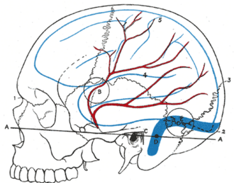
Branch of the maxillary artery that enters the skull through the foramen spinosum to supply the dura and skull. Tearing of its anterior branch under the pterion causes an epidural hematoma.
Core anatomy
- Location
Between the dura and the inner surface of the skull (extradural), in grooves on the temporal and parietal bones.
- Origin
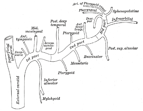
First part of the maxillary artery (a terminal branch of the external carotid artery), in the infratemporal fossa.
- Course
Passes between the two roots of the auriculotemporal nerve, enters the middle cranial fossa through the foramen spinosum, and divides into anterior (frontal) and posterior (parietal) branches. The anterior branch runs deep to the pterion.
- Branches
- Anterior (frontal) branch: crosses deep to the pterion toward the vertex
- Posterior (parietal) branch
- Small branches to the trigeminal ganglion, facial nerve and tympanic cavity
- Supplies
The dura mater and the bones of the cranial vault (not the brain).
- Anastomoses
With the ophthalmic artery (via the orbit), and with other meningeal arteries.
- Termination
Ends in the dura over the vault.
Found in
Mentioned in
Generated automatically from links in other entries.


Clinical correlations
Epidural (extradural) hematoma PANCE
- Mechanism / lesion
A temporal/parietal skull fracture (often at the pterion) tears the middle meningeal artery; arterial blood strips the dura from the skull.
- Classic presentation
Classic but inconsistent lucid interval after brief loss of consciousness, then headache, vomiting, decreasing consciousness, ipsilateral fixed dilated pupil (uncal herniation compressing CN III) and contralateral hemiparesis. CT: biconvex (lentiform) hyperdensity that does not cross suture lines. Treatment: urgent craniotomy and evacuation.
High-yield PANCE pearls
- Epidural = arterial (middle meningeal), biconvex, doesn't cross sutures, lucid interval.
- Subdural = venous (bridging veins), crescent, crosses sutures, elderly/alcohol use/anticoagulants.
- Middle meningeal artery enters through the foramen spinosum.
Imaging anatomy
- X-ray
Conventional angiography occasionally for traumatic pseudoaneurysm; embolization of the middle meningeal artery is also used to treat chronic subdural hematomas. verify
- CT
Non-contrast head CT: biconvex hyperdense extra-axial collection limited by sutures, often with an overlying fracture; "swirl sign" suggests active bleeding.
- Key views & landmarks
- Non-contrast head CT (brain and bone windows)
Physical exam
- Surface landmarks
The pterion lies about 3–4 cm above the midpoint of the zygomatic arch (two finger-breadths above the arch and a thumb's breadth behind the frontal process of the zygomatic bone).
- Strength / function testing
Serial Glasgow Coma Scale and pupil checks after head injury; a newly dilated pupil or falling GCS demands immediate CT and neurosurgery.
Self-quiz
The middle meningeal artery is a branch of which artery?
Correct answer: B
The middle meningeal artery arises from the first part of the maxillary artery (a terminal branch of the external carotid) and enters the skull through the foramen spinosum.
Show answer
B
The middle meningeal artery arises from the first part of the maxillary artery (a terminal branch of the external carotid) and enters the skull through the foramen spinosum.
Which CT finding best distinguishes an epidural from a subdural hematoma?
Correct answer: B
Epidural hematomas are biconvex and limited by suture lines, where the dura is firmly attached. Subdural hematomas are crescent-shaped and cross sutures (but not the midline falx).
Show answer
B
Epidural hematomas are biconvex and limited by suture lines, where the dura is firmly attached. Subdural hematomas are crescent-shaped and cross sutures (but not the midline falx).
Through which opening does the middle meningeal artery enter the cranial cavity?
Correct answer: D
The middle meningeal artery, from the maxillary artery, enters the middle cranial fossa through the foramen spinosum. The foramen ovale transmits V3 and the rotundum V2.
Show answer
D
The middle meningeal artery, from the maxillary artery, enters the middle cranial fossa through the foramen spinosum. The foramen ovale transmits V3 and the rotundum V2.
Which branch of the middle meningeal artery lies deep to the pterion?
Correct answer: B
The anterior (frontal) branch runs deep to the pterion, the thinnest part of the lateral skull, so a fracture there can tear it and cause an epidural hematoma.
Show answer
B
The anterior (frontal) branch runs deep to the pterion, the thinnest part of the lateral skull, so a fracture there can tear it and cause an epidural hematoma.
A patient with an expanding right epidural hematoma develops a fixed, dilated right pupil. What causes the pupil change?
Correct answer: C
An expanding hematoma pushes the uncus over the tentorium, compressing the ipsilateral oculomotor nerve; its superficial parasympathetic fibers are affected first, giving an ipsilateral fixed dilated pupil, often with contralateral hemiparesis.
Show answer
C
An expanding hematoma pushes the uncus over the tentorium, compressing the ipsilateral oculomotor nerve; its superficial parasympathetic fibers are affected first, giving an ipsilateral fixed dilated pupil, often with contralateral hemiparesis.
Flashcards
See also
- Skull
- External carotid artery
- Intracranial hemorrhage (comparison table)
- A lucid interval after a blow to the head (clinical case)