Vestibulocochlear nerve (CN VIII)
Special sensory nerve for hearing (cochlear part) and balance (vestibular part). It runs with CN VII through the internal acoustic meatus, where a vestibular schwannoma causes unilateral hearing loss.
Core anatomy
- Origin
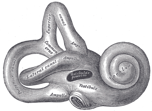
Cochlear part: bipolar neurons of the spiral ganglion in the cochlea. Vestibular part: bipolar neurons of the vestibular (Scarpa) ganglion from the semicircular canals, utricle and saccule (see inner ear).
- Course
Both parts pass through the internal acoustic meatus with the facial nerve and labyrinthine artery, cross the cerebellopontine angle and enter the brainstem at the pontomedullary junction. Cochlear fibers → cochlear nuclei → superior olive → lateral lemniscus → inferior colliculus → medial geniculate body (thalamus) → auditory cortex (superior temporal gyrus). Because of bilateral ascending connections, a unilateral lesion above the cochlear nuclei does not cause unilateral deafness. Vestibular fibers → vestibular nuclei → cerebellum, eye-movement nuclei (MLF — vestibulo-ocular reflex) and spinal cord (vestibulospinal tracts).
- Sensory
Hearing and balance (special somatic afferent).
- Relations
Cerebellopontine angle — CN V above, CN VII in front, the cerebellum and flocculus behind, CN IX–XI below.
Other structures innervated
Mentioned in
Generated automatically from links in other entries.
Swipe sideways to see the whole diagram →


Clinical correlations
Vestibular schwannoma (acoustic neuroma) PANCE
- Mechanism / lesion
Benign Schwann-cell tumor, usually of the vestibular division in the internal acoustic meatus/CPA; bilateral → neurofibromatosis type 2.
- Classic presentation
Unilateral (asymmetric) sensorineural hearing loss, tinnitus, imbalance; larger tumors compress CN V (reduced corneal reflex, facial numbness), CN VII, the cerebellum (ataxia) and cause hydrocephalus. Workup of asymmetric SNHL: audiogram then MRI with gadolinium of the internal auditory canals.
Peripheral vertigo PANCE
- Mechanism / lesion
Disorders of the labyrinth or vestibular nerve — BPPV (canaliths in the posterior canal), vestibular neuritis (viral, nerve only — no hearing loss), labyrinthitis (with hearing loss), Ménière disease (endolymphatic hydrops).
- Classic presentation
BPPV: seconds of vertigo with head position changes; Dix–Hallpike positive; Epley maneuver. Vestibular neuritis: days of continuous vertigo, nausea, horizontal nystagmus, normal hearing. Ménière: episodic vertigo (hours) + fluctuating low-frequency SNHL + tinnitus + aural fullness.
Central vs peripheral acute vestibular syndrome PANCE
- Mechanism / lesion
A cerebellar or brainstem stroke can mimic vestibular neuritis.
- Classic presentation
The HINTS exam (Head Impulse, Nystagmus, Test of Skew) in a patient with continuous vertigo and nystagmus: a normal head impulse, direction-changing or vertical nystagmus, or skew deviation suggests a central (stroke) cause → MRI.
High-yield PANCE pearls
- Unilateral SNHL + tinnitus → vestibular schwannoma → MRI of the IACs with contrast.
- Bilateral vestibular schwannomas → NF2.
- Brief positional vertigo → BPPV → Dix–Hallpike, Epley.
- Vertigo + fluctuating hearing loss + tinnitus + fullness → Ménière disease.
- Worrisome HINTS (normal head impulse, direction-changing nystagmus, skew) → stroke.
Imaging anatomy
- CT
Temporal bone CT for trauma and bony labyrinth disease.
- MRI
MRI with gadolinium of the internal auditory canals/CPA — vestibular schwannoma; MRI with DWI for suspected posterior circulation stroke (CT is insensitive in the posterior fossa).
Physical exam
- Strength / function testing
Whisper test or finger rub for hearing; Weber (tuning fork on the vertex — lateralizes to the affected ear in conductive loss, to the better ear in sensorineural loss) and Rinne (air > bone normally and in SNHL; bone > air in conductive loss) with a 512 Hz fork. Vestibular: nystagmus, head impulse test, Romberg, gait, Dix–Hallpike for positional vertigo.
Dix–Hallpike maneuver
- Technique
From sitting, turn the head 45° to one side and quickly lay the patient back with the head extended about 20° over the edge of the table; watch the eyes for up to 30 seconds.
- Positive test
After a short latency, upbeating torsional nystagmus toward the lower ear that fatigues within about 30–60 seconds, with vertigo.
- Significance
Posterior canal BPPV on the side of the lower ear → treat with the Epley repositioning maneuver.
Self-quiz
A 55-year-old has progressive hearing loss and ringing in the left ear. Weber lateralizes to the right, and Rinne is air > bone bilaterally. What is the next best test?
Correct answer: B
Weber to the right with air > bone indicates left sensorineural loss. Asymmetric SNHL with tinnitus needs MRI of the internal auditory canals to exclude a vestibular schwannoma.
Show answer
B
Weber to the right with air > bone indicates left sensorineural loss. Asymmetric SNHL with tinnitus needs MRI of the internal auditory canals to exclude a vestibular schwannoma.
A 62-year-old has vertigo for 30 seconds each time she rolls over in bed. Hearing is normal. Dix–Hallpike to the right produces upbeating torsional nystagmus that fades in 20 seconds. What is the treatment?
Correct answer: B
This is right posterior canal BPPV; the Epley canalith repositioning maneuver is curative in most patients. Long-term vestibular suppressants delay compensation.
Show answer
B
This is right posterior canal BPPV; the Epley canalith repositioning maneuver is curative in most patients. Long-term vestibular suppressants delay compensation.
Bilateral vestibular schwannomas are characteristic of which condition?
Correct answer: D
Bilateral vestibular schwannomas define neurofibromatosis type 2. Unilateral tumors usually occur sporadically.
Show answer
D
Bilateral vestibular schwannomas define neurofibromatosis type 2. Unilateral tumors usually occur sporadically.
Why does a unilateral lesion of the central auditory pathway above the cochlear nuclei not cause deafness in one ear?
Correct answer: B
Above the cochlear nuclei, auditory pathways ascend bilaterally (superior olive, lateral lemniscus), so a unilateral central lesion doesn't cause unilateral deafness. Unilateral hearing loss points to the cochlea, CN VIII or the cochlear nuclei.
Show answer
B
Above the cochlear nuclei, auditory pathways ascend bilaterally (superior olive, lateral lemniscus), so a unilateral central lesion doesn't cause unilateral deafness. Unilateral hearing loss points to the cochlea, CN VIII or the cochlear nuclei.
In a patient with continuous vertigo and nystagmus, which HINTS finding suggests a stroke rather than vestibular neuritis?
Correct answer: A
In vestibular neuritis the head impulse is abnormal (corrective saccade). A normal head impulse, direction-changing or vertical nystagmus, or skew deviation suggests a central (stroke) cause → MRI.
Show answer
A
In vestibular neuritis the head impulse is abnormal (corrective saccade). A normal head impulse, direction-changing or vertical nystagmus, or skew deviation suggests a central (stroke) cause → MRI.