Serratus anterior
Serrated muscle on the lateral thoracic wall that protracts the scapula, rotates it upward, and holds it against the chest wall.
Core anatomy
- Location
Lateral thoracic wall, forming the medial wall of the axilla. Lies deep to the scapula posteriorly.
- Group
Anterior axioappendicular muscles.
- Origin
External surfaces of the lateral parts of the upper 8–9 ribs by fleshy digitations. verify
- Insertion
Anterior (costal) surface of the medial border of the scapula, including the inferior angle.
- Innervation
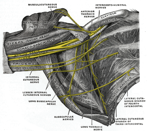
Long thoracic nerve (C5, C6, C7)
- Blood supply
Lateral thoracic and thoracodorsal arteries verify
- Action
- Protracts the scapula (drawing it anterolaterally, as when punching)
- Holds the scapula against the thoracic wall
- Rotates the scapula upward (inferior angle moves laterally), with trapezius, allowing the arm to be raised above 90°
- Relations
The long thoracic nerve runs on its superficial surface. The lower digitations interdigitate with the origin of the external oblique.
Forms a boundary of
Mentioned in
Generated automatically from links in other entries.

Clinical correlations
Winged scapula (serratus anterior paralysis) PANCE
- Mechanism / lesion
Injury to the long thoracic nerve: axillary dissection, mastectomy, chest tubes, blows to the lateral chest, heavy shoulder loads.
- Classic presentation
The medial border and inferior angle of the scapula project posteriorly, especially when pushing against a wall. The arm is hard to abduct above the horizontal.
High-yield PANCE pearls
- Serratus anterior: protraction + upward rotation of the scapula. Long thoracic nerve (C5–C7).
- Medial scapular winging after axillary surgery.
Imaging anatomy
- X-ray
The digitations are not visible. Chest radiographs are used for associated rib pathology.
- CT
Cross-sectional imaging shows a sheet of muscle along the lateral ribs with a "sawtooth" arrangement of digitations.
- MRI
Denervation edema or fatty atrophy in long thoracic nerve palsy.
- Ultrasound
Visible superficial to the ribs in the mid-axillary line, deep to latissimus dorsi. This plane is the target of the serratus anterior plane block used for chest wall analgesia (e.g. rib fractures).
Physical exam
- Surface landmarks
The digitations are visible and palpable on the lateral chest wall below the axilla in muscular people.
- Strength / function testing
Resisted protraction (pushing forward against the examiner's hand). Observe the scapula during wall push-ups.
Wall push-up test
- Technique
The patient pushes against a wall with the arms extended at shoulder height while the examiner watches the scapulae.
- Positive test
The medial border and inferior angle lift from the chest wall.
- Significance
Serratus anterior weakness (long thoracic nerve).
Self-quiz
A patient has difficulty raising the arm above the head after a lateral chest wall injury. Weakness of which scapular movement best explains this?
Correct answer: D
Serratus anterior (with trapezius) rotates the scapula upward so the glenoid faces superiorly, which is needed for abduction above 90°. The long thoracic nerve on the lateral chest wall is vulnerable to such injuries.
Show answer
D
Serratus anterior (with trapezius) rotates the scapula upward so the glenoid faces superiorly, which is needed for abduction above 90°. The long thoracic nerve on the lateral chest wall is vulnerable to such injuries.
Serratus anterior inserts on which part of the scapula?
Correct answer: C
Serratus anterior runs from the lateral ribs to the costal (anterior) surface of the medial border of the scapula. That's why its paralysis lets the medial border lift away from the chest wall.
Show answer
C
Serratus anterior runs from the lateral ribs to the costal (anterior) surface of the medial border of the scapula. That's why its paralysis lets the medial border lift away from the chest wall.
Which muscle forms most of the medial wall of the axilla?
Correct answer: C
The medial wall of the axilla is the thoracic wall covered by serratus anterior. Subscapularis forms most of the posterior wall and pectoralis major/minor the anterior wall.
Show answer
C
The medial wall of the axilla is the thoracic wall covered by serratus anterior. Subscapularis forms most of the posterior wall and pectoralis major/minor the anterior wall.
Serratus anterior produces which movement of the scapula, as when throwing a punch?
Correct answer: C
Serratus anterior protracts the scapula (draws it anterolaterally around the chest wall), holds it against the thorax and rotates it upward. Retraction is done mainly by the rhomboids and trapezius.
Show answer
C
Serratus anterior protracts the scapula (draws it anterolaterally around the chest wall), holds it against the thorax and rotates it upward. Retraction is done mainly by the rhomboids and trapezius.
Which muscle works with serratus anterior to rotate the scapula upward so the arm can be raised above 90°?
Correct answer: B
Trapezius (upper and lower fibres) and serratus anterior together rotate the scapula upward, turning the glenoid to face superiorly. The rhomboids, levator scapulae and pectoralis minor rotate it downward.
Show answer
B
Trapezius (upper and lower fibres) and serratus anterior together rotate the scapula upward, turning the glenoid to face superiorly. The rhomboids, levator scapulae and pectoralis minor rotate it downward.