Pancreas
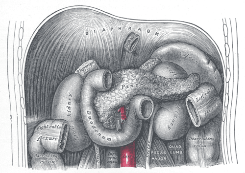
Mostly retroperitoneal gland lying across the posterior abdominal wall, with its head in the duodenal C-loop. Acute pancreatitis and pancreatic cancer are the key clinical problems.
Core anatomy
- Location
Retroperitoneal (except the tail), across the posterior abdominal wall at about L1–L2, behind the stomach and the lesser sac.
- Parts
- Head: within the C-loop of the duodenum; the uncinate process hooks behind the superior mesenteric vessels
- Neck: anterior to the confluence of the superior mesenteric and splenic veins, where the hepatic portal vein forms
- Body: crosses anterior to the aorta, the SMA origin and the left kidney
- Tail: runs in the splenorenal ligament to the splenic hilum (intraperitoneal; at risk during splenectomy)
- Main pancreatic duct (of Wirsung): joins the CBD at the hepatopancreatic ampulla → major duodenal papilla. Accessory duct (of Santorini): opens at the minor papilla
- Relations
Posterior: IVC, aorta, left renal vein, right crus of the diaphragm, left kidney and suprarenal gland. The splenic artery runs along its superior border and the splenic vein behind it. The CBD passes through or behind the head.
- Arterial supply
Head: superior pancreaticoduodenal arteries (from the gastroduodenal, celiac trunk) and inferior pancreaticoduodenal arteries (from the superior mesenteric artery), forming arcades. Neck, body, tail: branches of the splenic artery (dorsal, great and caudal pancreatic arteries).
- Venous drainage
Pancreatic veins to the splenic and superior mesenteric veins → portal vein.
- Lymphatic drainage
Pancreaticosplenic, pancreaticoduodenal, celiac and superior mesenteric nodes.
- Innervation
Vagus and sympathetic via the celiac and superior mesenteric plexuses. Visceral pain is epigastric and radiates to the back (it's retroperitoneal).
- Notes
Exocrine acini (digestive enzymes) make up most of the gland. Endocrine islets of Langerhans (insulin, glucagon, somatostatin) are most concentrated in the tail.
Mentioned in
- Celiac trunk
- Duodenum
- Gallbladder and biliary tree
- Hepatic portal vein
- Inferior vena cava
- Lesser sac
- Spleen
- Stomach
- Superior mesenteric artery
Generated automatically from links in other entries.
Swipe sideways to see the whole diagram →

Clinical correlations
Acute pancreatitis PANCE
- Mechanism / lesion
Premature activation of pancreatic enzymes. The most common causes are gallstones and alcohol; also hypertriglyceridemia (often above 1000 mg/dL), post-ERCP, hypercalcemia, medications and trauma.
- Classic presentation
Severe epigastric pain radiating to the back, often eased by leaning forward, with nausea and vomiting. Diagnosis needs 2 of 3: characteristic pain, lipase (or amylase) at least 3× the upper limit of normal, and characteristic imaging. Severe disease: SIRS, organ failure, Cullen/Grey Turner signs (hemorrhagic). Complications: necrosis, pseudocyst, ARDS, hypocalcemia.
Pancreatic adenocarcinoma PANCE
- Mechanism / lesion
Risk factors include smoking (the most important modifiable one), chronic pancreatitis, diabetes, obesity and familial syndromes. Most arise in the head.
- Classic presentation
Painless obstructive jaundice (head), weight loss, epigastric pain radiating to the back (body/tail), new-onset diabetes, Courvoisier sign, and Trousseau syndrome (migratory thrombophlebitis). CA 19-9 supports monitoring but isn't a screening test.
Chronic pancreatitis
- Mechanism / lesion
Recurrent inflammation and fibrosis, usually from long-term alcohol use.
- Classic presentation
Chronic epigastric pain, steatorrhea (exocrine insufficiency), diabetes (endocrine insufficiency), and pancreatic calcifications on imaging.
Pancreatic pseudocyst
- Mechanism / lesion
Encapsulated fluid collection without solid necrosis, usually 4 or more weeks after acute pancreatitis, often in the lesser sac.
- Classic presentation
Persistent pain, early satiety, epigastric mass. Many resolve; symptomatic ones are drained (often endoscopically).
High-yield PANCE pearls
- Acute pancreatitis: gallstones and alcohol are the top causes; diagnose with 2 of 3 (pain, lipase ≥ 3× ULN, imaging).
- Pain radiates to the back, relieved by leaning forward.
- Pancreatic head cancer: painless jaundice, Courvoisier sign, Trousseau syndrome; smoking is the main modifiable risk.
- Chronic pancreatitis triad: calcifications, steatorrhea, diabetes.
- Splenic artery along the superior border; SMV + splenic vein form the portal vein behind the neck.
Imaging anatomy
- X-ray
Chronic pancreatitis may show calcifications. A "sentinel loop" (localized ileus) in acute pancreatitis. Not diagnostic.
- CT
Contrast CT is used when the diagnosis is unclear or to assess complications (necrosis, collections), best performed after 48–72 hours. A pancreas-protocol CT stages cancer (vascular involvement determines resectability).
- MRI
MRCP shows ductal anatomy, CBD stones, and pancreas divisum.
- Ultrasound
Transabdominal ultrasound is done in every case of acute pancreatitis to look for gallstones. The pancreas itself is often obscured by bowel gas. Endoscopic ultrasound images small tumors and allows fine-needle biopsy.
- Key views & landmarks
- RUQ ultrasound (gallstones) in all acute pancreatitis
- Contrast CT (pancreas protocol for suspected cancer)
Physical exam
- Surface landmarks
Lies deep at about the transpyloric plane (L1), behind the stomach; the neck is roughly anterior to L1–L2 in the midline.
- Palpation
Epigastric tenderness and guarding in pancreatitis. A pseudocyst may form a palpable epigastric mass.
- Strength / function testing
Look for Cullen sign (periumbilical ecchymosis) and Grey Turner sign (flank ecchymosis) in severe hemorrhagic pancreatitis. Check for jaundice and a palpable gallbladder (Courvoisier). Hypocalcemia signs (Chvostek, Trousseau) in severe pancreatitis.
Self-quiz
A 45-year-old man has severe epigastric pain radiating to the back and vomiting after a weekend of heavy drinking. Lipase is 5 times the upper limit of normal. What is the most appropriate next step to look for an additional cause?
Correct answer: B
With characteristic pain and lipase at least 3× ULN, the diagnosis of acute pancreatitis is made without CT. RUQ ultrasound is recommended in all patients to look for gallstones as a cause.
Show answer
B
With characteristic pain and lipase at least 3× ULN, the diagnosis of acute pancreatitis is made without CT. RUQ ultrasound is recommended in all patients to look for gallstones as a cause.
A 70-year-old smoker has painless jaundice, weight loss, and a palpable non-tender gallbladder. Where is the tumor most likely located?
Correct answer: C
A tumor in the head of the pancreas compresses the distal CBD, causing painless obstructive jaundice and Courvoisier sign.
Show answer
C
A tumor in the head of the pancreas compresses the distal CBD, causing painless obstructive jaundice and Courvoisier sign.
Which part of the pancreas lies in the splenorenal ligament and can be injured during splenectomy?
Correct answer: E
The tail runs in the splenorenal ligament to the splenic hilum; injury during splenectomy can cause a pancreatic fistula.
Show answer
E
The tail runs in the splenorenal ligament to the splenic hilum; injury during splenectomy can cause a pancreatic fistula.
The hepatic portal vein is formed behind which part of the pancreas?
Correct answer: B
The superior mesenteric vein and splenic vein unite behind the neck of the pancreas to form the portal vein.
Show answer
B
The superior mesenteric vein and splenic vein unite behind the neck of the pancreas to form the portal vein.
Which combination is the classic triad of chronic pancreatitis?
Correct answer: E
Chronic pancreatitis (usually from long-term alcohol use) causes calcifications, steatorrhea (exocrine insufficiency) and diabetes (endocrine insufficiency), with chronic epigastric pain. The other choices describe cholangitis, pancreatic head cancer, acute pancreatitis criteria and tamponade.
Show answer
E
Chronic pancreatitis (usually from long-term alcohol use) causes calcifications, steatorrhea (exocrine insufficiency) and diabetes (endocrine insufficiency), with chronic epigastric pain. The other choices describe cholangitis, pancreatic head cancer, acute pancreatitis criteria and tamponade.