Psoas major
Long muscle beside the lumbar spine that joins iliacus to flex the hip. The lumbar plexus forms within it, and the psoas sign reflects inflammation of structures lying on it.
Core anatomy
- Location
Posterior abdominal wall, beside the lumbar vertebral bodies, passing under the inguinal ligament into the thigh.
- Group
Posterior abdominal wall muscles (with iliacus and quadratus lumborum). Psoas + iliacus = iliopsoas.
- Origin
Sides of the vertebral bodies of T12–L5 and the intervening discs; transverse processes of the lumbar vertebrae.
- Insertion
Lesser trochanter of the femur (via a common tendon with iliacus).
- Innervation
Anterior rami of lumbar nerves L1–L3 (from the lumbar plexus)
- Blood supply
Lumbar arteries, iliolumbar artery (psoas branch) and branches of the external iliac/femoral arteries.
- Action
- Flexes the thigh at the hip (with iliacus), the main hip flexor
- Flexes the trunk on the thigh (sitting up from supine) and flexes the lumbar spine laterally
- Relations
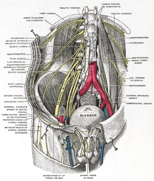
The lumbar plexus forms within the muscle: the femoral nerve emerges from its lateral border, the obturator nerve from its medial border, and the genitofemoral nerve from its anterior surface. The ureter and gonadal vessels descend on its anterior surface. The kidney lies on its upper part.
Mentioned in
Generated automatically from links in other entries.

Clinical correlations
Psoas abscess PANCE
- Mechanism / lesion
Primary: hematogenous spread (Staphylococcus aureus, often in people who inject drugs, immunocompromised). Secondary: spread from adjacent disease such as vertebral osteomyelitis/TB (Pott disease), Crohn disease, diverticulitis or appendicitis.
- Classic presentation
Fever, back or flank pain, limp, and the hip held flexed; pain on hip extension (psoas sign). CT confirms it; treated with drainage and antibiotics.
Psoas sign in appendicitis PANCE
- Mechanism / lesion
An inflamed retrocecal appendix lies against the right psoas.
- Classic presentation
RLQ pain on passive extension of the right hip or on active flexion against resistance.
Retroperitoneal (psoas) hematoma
- Mechanism / lesion
Bleeding in anticoagulated patients or after femoral arterial access.
- Classic presentation
Flank or groin pain, hip flexion, falling hemoglobin, and femoral nerve palsy (weak knee extension, reduced patellar reflex, anterior thigh numbness).
High-yield PANCE pearls
- Psoas = main hip flexor; inserts on the lesser trochanter; L1–L3.
- Psoas sign → retrocecal appendicitis or psoas abscess.
- Psoas abscess: fever, back pain, limp, flexed hip; think S. aureus, Pott disease, Crohn.
- Femoral nerve emerges from its lateral border; obturator from its medial border.
Imaging anatomy
- X-ray
Loss of the psoas shadow on an abdominal film is a nonspecific sign of retroperitoneal disease.
- CT
CT with contrast is the test of choice for psoas abscess or hematoma (a rim-enhancing collection within an enlarged muscle).
- MRI
Best for associated spinal osteomyelitis/discitis.
- Key views & landmarks
- CT abdomen/pelvis with contrast
Physical exam
- Surface landmarks
Not palpable directly; its tendon passes deep to the inguinal ligament lateral to the femoral vessels.
- Strength / function testing
Resisted hip flexion in sitting (L2–L3 myotome).
Psoas sign
- Technique
With the patient lying on the left side, passively extend the right hip; or have the supine patient flex the right hip against resistance.
- Positive test
Lower abdominal or flank pain.
- Significance
Irritation of the psoas (retrocecal appendicitis, psoas abscess or hematoma).
Self-quiz
A 45-year-old man who injects drugs has fever, right flank pain, and a limp. He holds his right hip slightly flexed, and extending it is painful. What is the most likely diagnosis?
Correct answer: B
Fever, back/flank pain and a flexed hip with pain on extension suggest a psoas abscess, often S. aureus in people who inject drugs. CT confirms it.
Show answer
B
Fever, back/flank pain and a flexed hip with pain on extension suggest a psoas abscess, often S. aureus in people who inject drugs. CT confirms it.
Where does psoas major insert?
Correct answer: B
Psoas major and iliacus insert together on the lesser trochanter of the femur.
Show answer
B
Psoas major and iliacus insert together on the lesser trochanter of the femur.
Which nerve emerges from the lateral border of psoas major?
Correct answer: C
The femoral nerve emerges laterally, the obturator medially, and the genitofemoral nerve through its anterior surface.
Show answer
C
The femoral nerve emerges laterally, the obturator medially, and the genitofemoral nerve through its anterior surface.