Cervical lymph nodes
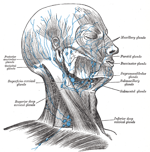
Superficial and deep nodes of the head and neck, grouped clinically into levels I–VI. Lymphadenopathy is common, and a persistent neck mass in an adult is cancer until proven otherwise.
Core anatomy
- Location
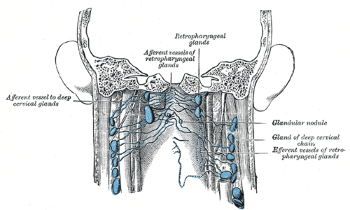
Superficial ring at the junction of head and neck (occipital, mastoid, preauricular/parotid, submandibular, submental) and superficial cervical nodes along the external jugular vein; the deep cervical chain along the internal jugular vein under sternocleidomastoid. Key named nodes — jugulodigastric (tonsil, below the angle of the jaw) and jugulo-omohyoid (tongue).
- Drains
All lymph from the head and neck ultimately reaches the deep cervical chain. Tip of tongue and lower lip → submental. Face, lateral tongue → submandibular. Scalp, ear → occipital/mastoid/preauricular. Tonsil, pharynx → jugulodigastric. Thyroid and larynx → central compartment and deep nodes.
- Drains to
Deep cervical nodes → jugular trunk → on the left the thoracic duct, on the right the right lymphatic duct → the junction of the internal jugular and subclavian veins.
- Relations
Clinical levels: I submental/submandibular; II upper jugular (skull base to hyoid); III middle jugular (hyoid to cricoid); IV lower jugular (cricoid to clavicle); V posterior triangle; VI central compartment (pretracheal, paratracheal, prelaryngeal). VII superior mediastinum in some systems. The spinal accessory nerve runs through level V (see triangles of the neck).
- Notes
The left supraclavicular node (Virchow) receives lymph from the thoracic duct region and can enlarge with abdominal or thoracic cancers (Troisier sign).
Mentioned in
- Internal jugular vein
- Palatine tonsils
- Parathyroid glands
- Parotid gland
- Pharynx
- Scalp
- Submandibular gland
- Thyroid gland
- Tongue
Generated automatically from links in other entries.


Clinical correlations
Cervical lymphadenopathy PANCE
- Mechanism / lesion
Usually reactive (viral URI, strep pharyngitis, dental infection); also EBV/CMV, cat-scratch disease (Bartonella), TB (scrofula), lymphoma and metastatic cancer.
- Classic presentation
Reactive nodes are tender, soft, mobile and under about 1 cm. Concerning: over 1.5–2 cm, hard, fixed, non-tender, supraclavicular location, persistent over 2–4 weeks, weight loss/night sweats/fever, age over 40, smoking or alcohol history. Posterior cervical nodes suggest mononucleosis; anterior nodes, strep.
Adult neck mass PANCE
- Mechanism / lesion
In adults a persistent lateral neck mass is metastatic squamous cell carcinoma (oral cavity, oropharynx — often HPV, larynx) or lymphoma until proven otherwise.
- Classic presentation
Workup: full head and neck exam with laryngoscopy, contrast CT of the neck, and fine-needle aspiration (avoid open excision first). HPV-related oropharyngeal cancer often presents as a cystic node.
Virchow node PANCE
- Mechanism / lesion
Metastasis to the left supraclavicular node via the thoracic duct.
- Classic presentation
A hard left supraclavicular node → look for gastric and other abdominal or thoracic cancers. Right supraclavicular nodes drain the mediastinum, lungs and esophagus.
Lymphoma
- Mechanism / lesion
Hodgkin (Reed–Sternberg cells; young adults; contiguous spread) or non-Hodgkin lymphoma.
- Classic presentation
Painless, rubbery cervical or supraclavicular nodes ± B symptoms; Hodgkin nodes may hurt after alcohol. Diagnosis requires excisional biopsy (architecture), unlike SCC.
High-yield PANCE pearls
- Hard, fixed, painless, > 1.5 cm or supraclavicular node → biopsy/FNA.
- Left supraclavicular (Virchow) node → gastric/abdominal cancer.
- Adult with persistent neck mass → head and neck SCC until proven otherwise → CT + FNA + scope.
- Posterior cervical nodes → mono; anterior → strep.
- Suspected lymphoma → excisional biopsy (FNA is insufficient).
Imaging anatomy
- CT
Contrast CT of the neck for adult neck masses and cancer staging; necrotic nodes suggest SCC or TB.
- MRI
Complementary for staging; PET-CT for staging lymphoma and head and neck cancers or finding an unknown primary.
- Ultrasound
First-line for superficial nodes in children and to guide FNA; features of malignancy — round shape, loss of the fatty hilum, necrosis.
- Key views & landmarks
- Contrast CT neck
- Ultrasound-guided FNA
Physical exam
- Surface landmarks
Levels I–VI as above; the supraclavicular fossa behind the clavicle.
- Palpation
Use a systematic sequence with the pads of the fingers, patient's neck slightly flexed: preauricular, posterior auricular, occipital, tonsillar (jugulodigastric), submandibular, submental, superficial cervical, deep cervical chain (hook around SCM), posterior cervical, supraclavicular (have the patient shrug or perform Valsalva). Note size, consistency, tenderness, mobility and matting.
Self-quiz
A 58-year-old man who smokes has a painless, firm 3-cm mass in the left upper neck for 6 weeks. There are no infectious symptoms. What is the most appropriate next step?
Correct answer: C
A persistent neck mass in an adult smoker is metastatic SCC until proven otherwise. Look for the primary (endoscopy), image (CT) and obtain cytology (FNA). Open excisional biopsy first can seed the neck and compromise treatment.
Show answer
C
A persistent neck mass in an adult smoker is metastatic SCC until proven otherwise. Look for the primary (endoscopy), image (CT) and obtain cytology (FNA). Open excisional biopsy first can seed the neck and compromise treatment.
A hard left supraclavicular lymph node is most classically associated with which cancer?
Correct answer: B
Virchow node (left supraclavicular) receives lymph near the termination of the thoracic duct and is a classic sign of gastric (and other abdominal) cancer.
Show answer
B
Virchow node (left supraclavicular) receives lymph near the termination of the thoracic duct and is a classic sign of gastric (and other abdominal) cancer.
When lymphoma is suspected in an enlarged cervical node, which biopsy is needed?
Correct answer: A
Lymphoma diagnosis needs the node architecture, so an excisional biopsy is required; FNA is insufficient. For a suspected metastatic squamous cell carcinoma, FNA is preferred and open excision is avoided first.
Show answer
A
Lymphoma diagnosis needs the node architecture, so an excisional biopsy is required; FNA is insufficient. For a suspected metastatic squamous cell carcinoma, FNA is preferred and open excision is avoided first.
Lymph from the palatine tonsil drains first to which named node?
Correct answer: D
The tonsil drains to the jugulodigastric (tonsillar) node of the deep cervical chain, just below the angle of the mandible, which enlarges in tonsillitis. The jugulo-omohyoid node is associated with the tongue.
Show answer
D
The tonsil drains to the jugulodigastric (tonsillar) node of the deep cervical chain, just below the angle of the mandible, which enlarges in tonsillitis. The jugulo-omohyoid node is associated with the tongue.
A teenager with pharyngitis has prominent posterior cervical lymphadenopathy. Which diagnosis does this pattern suggest?
Correct answer: B
Posterior cervical nodes suggest infectious mononucleosis (EBV); anterior cervical nodes suggest streptococcal pharyngitis.
Show answer
B
Posterior cervical nodes suggest infectious mononucleosis (EBV); anterior cervical nodes suggest streptococcal pharyngitis.