Sacral plexus
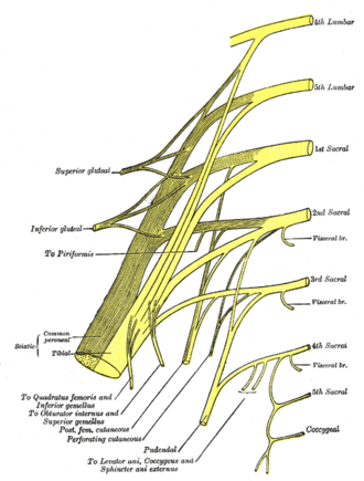
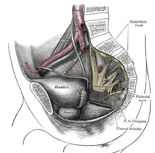
Nerve network on the posterior pelvic wall (L4–S4) that gives the sciatic, gluteal and pudendal nerves. It supplies the buttock, posterior thigh, nearly all of the leg and foot, the pelvic floor and the perineum.
Core anatomy
- Location
On the anterior surface of piriformis on the posterior pelvic wall, behind the internal iliac vessels and the ureter.
- Root values
Lumbosacral trunk (part of L4 plus L5, joining from the lumbar plexus over the pelvic brim) and the ventral rami of S1–S4.
- Origin
Ventral rami of L4–S4.
- Course
The rami converge toward the greater sciatic foramen; most branches leave the pelvis through it, above or below piriformis.
- Branches
- Sciatic nerve (L4–S3): the largest nerve in the body; leaves below piriformis (usually), supplies the posterior thigh and, through the tibial and common fibular nerves, all muscles below the knee
- Superior gluteal nerve (L4–S1): leaves above piriformis; supplies gluteus medius, gluteus minimus and tensor fasciae latae
- Inferior gluteal nerve (L5–S2): below piriformis; supplies gluteus maximus
- Posterior femoral cutaneous nerve (S1–S3): skin of the posterior thigh and part of the perineum
- Pudendal nerve (S2–S4): perineum
- Nerve to obturator internus (and superior gemellus) (L5–S2) and nerve to quadratus femoris (and inferior gemellus) (L4–S1)
- Nerve to piriformis (S1–S2); nerves to levator ani and coccygeus (S3–S4)
- Pelvic splanchnic nerves (S2–S4): parasympathetic (see pelvic autonomic nerves)
- Motor
Gluteal muscles, the lateral rotators of the hip, hamstrings, all muscles of the leg and foot, the pelvic floor and the perineal muscles.
- Sensory
Posterior thigh, most of the leg and foot (except the medial side supplied by the saphenous nerve), the buttock (partly) and the perineum.
- Relations
The internal iliac vessels and their branches weave between the roots (the superior gluteal artery usually passes between L4/5 and S1, the inferior gluteal between S1/2 or S2/3). The rectum and sigmoid colon lie in front.
Muscles innervated
- Gluteus maximus — Inferior gluteal nerve (L5–S2), from the sacral plexus
- Gluteus medius and minimus — Superior gluteal nerve (L4–S1), from the sacral plexus
- Piriformis — Nerve to piriformis (S1–S2), from the sacral plexus
Muscles innervated via branches
- Adductor muscles of the thigh — via Sciatic nerve
- Hamstrings — via Sciatic nerve
Branches
Mentioned in
Generated automatically from links in other entries.


Clinical correlations
Superior gluteal nerve injury (Trendelenburg sign) PANCE
- Mechanism / lesion
Injury from a misplaced intramuscular injection, hip surgery, or pelvic fracture weakens gluteus medius and minimus.
- Classic presentation
When standing on the affected leg, the opposite side of the pelvis drops (positive Trendelenburg sign), with a waddling or lurching (compensated Trendelenburg) gait.
Sciatic nerve injury from intramuscular injection PANCE
- Mechanism / lesion
Injection into the lower or medial buttock can hit the sciatic nerve as it leaves below piriformis.
- Classic presentation
Pain and weakness below the knee (foot drop is common because the fibular division is most vulnerable). Give gluteal injections in the upper outer quadrant (or the ventrogluteal site).
Lumbosacral plexopathy
- Mechanism / lesion
Compression or infiltration by pelvic tumors (rectal, cervical, prostate), retroperitoneal hematoma, radiation, diabetic amyotrophy, or pressure from the fetal head during labor.
- Classic presentation
Leg pain and weakness in more than one root or nerve distribution, with preserved paraspinal muscles on EMG (distinguishing it from radiculopathy).
High-yield PANCE pearls
- Positive Trendelenburg = superior gluteal nerve (gluteus medius/minimus) weakness; the opposite hip drops.
- Superior gluteal nerve and vessels are the only structures above piriformis.
- IM gluteal injections go in the upper outer quadrant to avoid the sciatic nerve.
- Sacral plexus roots: L4–S4 (lumbosacral trunk + S1–S4).
Imaging anatomy
- CT
CT shows sacral fractures (with neural foramen involvement) and pelvic masses.
- MRI
MRI of the lumbosacral plexus (or MR neurography) shows tumor infiltration, hematoma, radiation change and nerve enlargement.
- Key views & landmarks
- MRI lumbosacral plexus (coronal STIR/T1)
Physical exam
- Surface landmarks
The sciatic nerve emerges from under piriformis at about the midpoint of a line from the PSIS to the ischial tuberosity, then descends between the greater trochanter and ischial tuberosity.
- Strength / function testing
Myotomes: hip abduction (L5, superior gluteal), hip extension (S1, inferior gluteal), knee flexion (L5–S1), ankle dorsiflexion (L4–L5), great toe extension (L5), plantarflexion (S1). Ankle reflex (S1). Sensation over the posterior thigh, lateral leg and foot.
Trendelenburg test
- Technique
The patient stands on one leg (the tested side) with the other hip and knee flexed, for about 30 seconds.
- Positive test
The pelvis drops on the side of the lifted leg, or the patient leans the trunk over the stance leg.
- Significance
Weakness of the hip abductors (gluteus medius/minimus) on the stance side — superior gluteal nerve injury, L5 radiculopathy or hip pathology.
Self-quiz
After an intramuscular injection into the buttock, a patient walks with a lurching gait. When standing on the right leg, the left side of the pelvis drops. Which nerve is most likely injured?
Correct answer: B
When standing on the right leg, the right gluteus medius and minimus must hold the pelvis level. Weakness (right superior gluteal nerve) lets the unsupported left side drop — a positive Trendelenburg sign.
Show answer
B
When standing on the right leg, the right gluteus medius and minimus must hold the pelvis level. Weakness (right superior gluteal nerve) lets the unsupported left side drop — a positive Trendelenburg sign.
Which nerve supplies gluteus maximus?
Correct answer: B
The inferior gluteal nerve (L5–S2) supplies gluteus maximus, the main hip extensor used to rise from a chair or climb stairs. The superior gluteal nerve supplies gluteus medius, minimus and tensor fasciae latae.
Show answer
B
The inferior gluteal nerve (L5–S2) supplies gluteus maximus, the main hip extensor used to rise from a chair or climb stairs. The superior gluteal nerve supplies gluteus medius, minimus and tensor fasciae latae.
Which structure passes through the greater sciatic foramen above the piriformis muscle?
Correct answer: C
Only the superior gluteal nerve and vessels pass above piriformis; everything else from the sacral plexus passes below it.
Show answer
C
Only the superior gluteal nerve and vessels pass above piriformis; everything else from the sacral plexus passes below it.
Intramuscular gluteal injections are given in the upper outer quadrant mainly to avoid which nerve?
Correct answer: C
Injections into the lower or medial buttock can hit the sciatic nerve as it leaves below piriformis, causing pain and weakness below the knee (often foot drop, because the fibular division is most vulnerable). Use the upper outer quadrant or the ventrogluteal site.
Show answer
C
Injections into the lower or medial buttock can hit the sciatic nerve as it leaves below piriformis, causing pain and weakness below the knee (often foot drop, because the fibular division is most vulnerable). Use the upper outer quadrant or the ventrogluteal site.
The lumbosacral trunk that joins the sacral plexus is formed from which ventral rami?
Correct answer: A
The lumbosacral trunk (part of L4 plus L5) descends over the pelvic brim from the lumbar plexus to join the ventral rami of S1–S4, so the sacral plexus roots are L4–S4.
Show answer
A
The lumbosacral trunk (part of L4 plus L5) descends over the pelvic brim from the lumbar plexus to join the ventral rami of S1–S4, so the sacral plexus roots are L4–S4.