Median nerve
Formed by roots from the lateral and medial cords. It supplies most of the anterior forearm and the thenar muscles, and passes through the carpal tunnel.
Core anatomy
- Location
Arm (medial) → cubital fossa → anterior forearm (between the flexor layers) → carpal tunnel → palm.
- Root values
C6, C7, C8, T1 (± C5) verify
- Origin
Lateral root from the lateral cord and medial root from the medial cord of the brachial plexus. The roots unite anterior to the third part of the axillary artery.
- Course
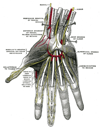
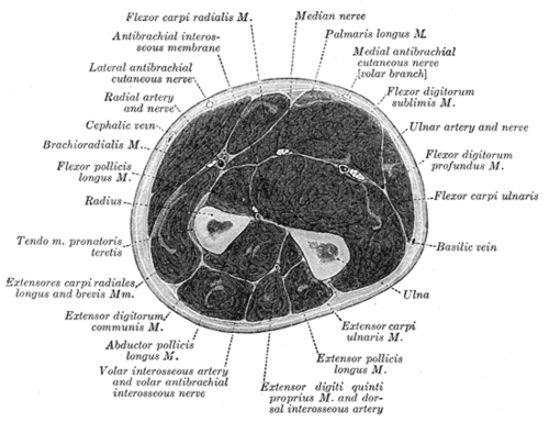
Arm: descends alongside the brachial artery, lateral to it proximally, then crosses (usually anteriorly) to lie medial to it at the elbow. It gives no named muscular branches in the arm. Cubital fossa: the most medial of the main contents, medial to the brachial artery and deep to the bicipital aponeurosis. Forearm: enters between the two heads of pronator teres and gives off the anterior interosseous nerve (AIN). It then runs between flexor digitorum superficialis (FDS) and flexor digitorum profundus (FDP), adherent to the deep surface of FDS. Wrist: becomes superficial between the tendons of FCR and FDS, deep to palmaris longus. The palmar cutaneous branch arises proximal to the flexor retinaculum and passes superficial to it. The nerve then passes through the carpal tunnel deep to the flexor retinaculum and divides into the recurrent (thenar motor) branch and common palmar digital nerves.
- Branches
- Muscular (forearm): Pronator teres, flexor carpi radialis, palmaris longus, flexor digitorum superficialis
- Anterior interosseous nerve: flexor pollicis longus, lateral half of FDP (digits 2–3), pronator quadratus (no cutaneous branch)
- Palmar cutaneous branch: lateral palm; arises proximal to and passes superficial to the flexor retinaculum
- Recurrent (thenar) branch: abductor pollicis brevis, opponens pollicis, superficial head of flexor pollicis brevis
- Common/proper palmar digital nerves: lateral two lumbricals; skin of the lateral 3½ digits
- Motor
Most of the anterior forearm flexors except FCU and the medial half of FDP (ulnar). In the hand: "LOAF", the lateral two Lumbricals, Opponens pollicis, Abductor pollicis brevis and Flexor pollicis brevis (superficial head).
- Sensory
Palmar surface of the lateral 3½ digits (thumb, index, middle, lateral half of ring) and the dorsal distal phalanges/nail beds of those digits. Lateral palm via the palmar cutaneous branch. Most reliable test site: the pad of the index finger.
- Relations
Crosses the brachial artery in the arm. In the carpal tunnel it is the most superficial structure, lying with the nine long flexor tendons.
Muscles innervated
- Pronator teres — median nerve (C6, C7)
Other structures innervated
Found in
Mentioned in
Generated automatically from links in other entries.
Swipe sideways to see the whole diagram →


Clinical correlations
Carpal tunnel syndrome PANCE
- Mechanism / lesion
Compression of the median nerve in the carpal tunnel. It is the most common entrapment neuropathy. Associations: pregnancy, hypothyroidism, diabetes, rheumatoid arthritis, obesity, acromegaly, dialysis-related amyloidosis, repetitive wrist use, and space-occupying lesions (ganglion, lunate dislocation).
- Classic presentation
Paresthesias and pain in the lateral 3½ digits, worse at night, often relieved by shaking the hand ("flick sign"). Later: weakness of thumb abduction/opposition and thenar atrophy. Sensation over the thenar eminence is spared because the palmar cutaneous branch passes superficial to the retinaculum.
Anterior interosseous nerve palsy PANCE
- Mechanism / lesion
Injury in the proximal forearm. Classically associated with pediatric supracondylar humerus fractures (the AIN is the nerve most often injured in extension-type fractures), or with compression or neuritis. verify
- Classic presentation
Can't make an "OK" sign: the thumb IP joint and index DIP joint don't flex (FPL and FDP to the index), so the pinch is flat. Weak pronation with the elbow flexed (pronator quadratus). No sensory loss.
Proximal median nerve lesion ("hand of benediction") PANCE
- Mechanism / lesion
Injury at or above the elbow, e.g. supracondylar fracture or penetrating trauma.
- Classic presentation
When the patient tries to make a fist, the index and middle fingers stay extended (loss of FDS and the lateral FDP) while the ring and little fingers flex (ulnar FDP). This is the "hand of benediction". There is also thenar weakness and median sensory loss including the thenar eminence.
Pronator syndrome
- Mechanism / lesion
Compression near the elbow: between the heads of pronator teres, under the bicipital aponeurosis, or under the FDS arch.
- Classic presentation
Aching of the volar forearm with median-distribution paresthesias. Unlike carpal tunnel syndrome, sensation over the thenar eminence is reduced (palmar cutaneous branch involved) and night symptoms are less prominent.
Laceration at the wrist ("ape hand")
- Mechanism / lesion
A cut at the wrist crease (e.g. glass or a suicide attempt) divides the nerve distal to its forearm branches.
- Classic presentation
Thenar paralysis and atrophy leave the thumb lying adducted in the plane of the palm ("ape hand"), unable to oppose. Median sensory loss in the digits. Forearm flexors are intact.
High-yield PANCE pearls
- Carpal tunnel syndrome: nocturnal paresthesias of the lateral 3½ digits; Tinel, Phalen, and the carpal compression (Durkan) test; confirm with nerve conduction studies/EMG.
- Associations with CTS: pregnancy, hypothyroidism, diabetes, RA, acromegaly.
- Initial management of mild–moderate CTS commonly includes a wrist splint in neutral (especially at night). Thenar atrophy or severe/progressive deficit prompts surgical referral.
- Supracondylar fracture (child) + can't make an "OK" sign → AIN injury.
- Thenar sensation spared in CTS (palmar cutaneous branch); reduced in pronator syndrome.
Imaging anatomy
- X-ray
Not visible. A carpal tunnel (axial) view can show bony causes of compression. At the elbow, radiographs evaluate supracondylar fractures: look for the posterior fat pad sign and an abnormal anterior humeral line.
- MRI
In carpal tunnel syndrome: enlargement of the nerve at the pisiform level, flattening at the hamate level, increased T2 signal, and palmar bowing of the flexor retinaculum. MRI also shows masses and tenosynovitis.
- Ultrasound
First-line imaging adjunct for carpal tunnel syndrome. The nerve appears as a honeycomb (fascicular) oval structure beneath the flexor retinaculum. An increased cross-sectional area at the tunnel inlet (pisiform level) supports the diagnosis. verify It also detects anatomic variants (bifid median nerve, persistent median artery) and masses.
- Key views & landmarks
- Ultrasound transverse at pisiform (inlet) and hook of hamate (outlet) levels
- Elbow lateral radiograph for pediatric supracondylar fracture
Physical exam
- Surface landmarks
At the wrist, the nerve lies deep to palmaris longus, between the FCR and FDS tendons, just proximal to the distal wrist crease. (Palmaris longus is absent in some people.)
- Palpation
The nerve itself is not reliably palpable; percussion over the carpal tunnel is used for the Tinel sign.
- Strength / function testing
Thumb abduction against resistance (APB), the most reliable median motor test in the hand; opposition. "OK" sign for the AIN (FPL + FDP index). Fist making: look for a hand of benediction. Two-point discrimination or light touch on the pad of the index finger.
Tinel sign (wrist)
- Technique
Tap over the median nerve at the carpal tunnel.
- Positive test
Tingling radiating into the median-innervated digits.
- Significance
Suggests carpal tunnel syndrome. Sensitivity and specificity are moderate.
Phalen test
- Technique
Hold the wrists in full flexion (dorsal surfaces of the hands pressed together) for 60 seconds.
- Positive test
Paresthesias in the median distribution.
- Significance
Suggests carpal tunnel syndrome.
Carpal compression test (Durkan)
- Technique
The examiner presses with the thumbs directly over the carpal tunnel for up to 30 seconds.
- Positive test
Paresthesias in the median distribution.
- Significance
Suggests carpal tunnel syndrome. Often reported as the most sensitive of the provocative tests.
Self-quiz
A 29-year-old woman at 32 weeks' gestation reports numbness of the right thumb, index and middle fingers that wakes her at night and improves when she shakes her hand. Which finding would suggest the lesion is proximal to the carpal tunnel rather than within it?
Correct answer: C
The palmar cutaneous branch arises proximal to the flexor retinaculum and passes superficial to it, so thenar skin sensation is spared in carpal tunnel syndrome. Thenar sensory loss points to a more proximal lesion such as pronator syndrome. The other findings occur in carpal tunnel syndrome.
Show answer
C
The palmar cutaneous branch arises proximal to the flexor retinaculum and passes superficial to it, so thenar skin sensation is spared in carpal tunnel syndrome. Thenar sensory loss points to a more proximal lesion such as pronator syndrome. The other findings occur in carpal tunnel syndrome.
A 7-year-old boy falls from monkey bars onto an outstretched hand and sustains an extension-type supracondylar humerus fracture. After reduction he can't flex the IP joint of the thumb or the DIP joint of the index finger. Sensation in the hand is normal. Which nerve is injured?
Correct answer: E
The AIN supplies FPL, the lateral FDP and pronator quadratus and has no cutaneous branch. The patient can't form an "OK" sign. It is the nerve most often injured in extension-type supracondylar fractures.
Show answer
E
The AIN supplies FPL, the lateral FDP and pronator quadratus and has no cutaneous branch. The patient can't form an "OK" sign. It is the nerve most often injured in extension-type supracondylar fractures.
Which intrinsic hand muscle is NOT innervated by the median nerve?
Correct answer: C
Adductor pollicis is supplied by the deep branch of the ulnar nerve (that's the basis of the Froment sign). The median nerve supplies "LOAF": lateral two lumbricals, opponens pollicis, abductor pollicis brevis and the superficial head of flexor pollicis brevis.
Show answer
C
Adductor pollicis is supplied by the deep branch of the ulnar nerve (that's the basis of the Froment sign). The median nerve supplies "LOAF": lateral two lumbricals, opponens pollicis, abductor pollicis brevis and the superficial head of flexor pollicis brevis.
A patient tries to make a fist, but the index and middle fingers remain extended. Where is the lesion?
Correct answer: B
The hand of benediction results from loss of FDS and the lateral half of FDP, which are supplied in the forearm. The lesion must therefore be proximal to those branches, at or above the elbow. A lesion at the wrist spares the long flexors.
Show answer
B
The hand of benediction results from loss of FDS and the lateral half of FDP, which are supplied in the forearm. The lesion must therefore be proximal to those branches, at or above the elbow. A lesion at the wrist spares the long flexors.
A man cuts his wrist crease on broken glass and divides the median nerve at that level. Which function is preserved?
Correct answer: A
A wrist-level laceration is distal to the forearm branches, including the anterior interosseous nerve, so FDP to the index (DIP flexion), FPL and the forearm flexors still work. The thenar muscles and median digital sensation are lost, leaving an adducted, unopposable thumb ("ape hand").
Show answer
A
A wrist-level laceration is distal to the forearm branches, including the anterior interosseous nerve, so FDP to the index (DIP flexion), FPL and the forearm flexors still work. The thenar muscles and median digital sensation are lost, leaving an adducted, unopposable thumb ("ape hand").
Flashcards
See also
- Carpal tunnel
- Cubital fossa
- Upper limb nerve injuries (comparison table)
- Numb fingers that wake her at night (clinical case)