Thoracic aorta
The aorta within the thorax (ascending aorta, arch and descending thoracic aorta). It's the site of dissection, coarctation, traumatic injury and aneurysm.
Core anatomy
- Location
Middle mediastinum (ascending), superior mediastinum (arch), posterior mediastinum (descending).
- Origin
Aortic orifice of the left ventricle, above the aortic valve.
- Course
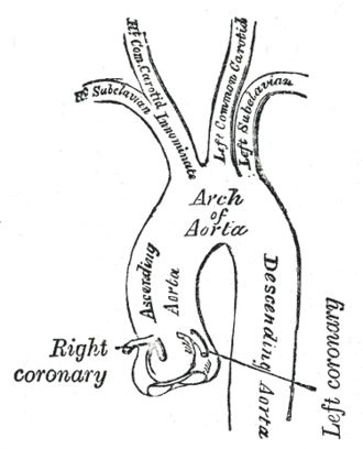
Ascending aorta: from the aortic valve, within the pericardium, rising to the level of the sternal angle. Arch: begins and ends at the sternal angle (T4/T5), passing posteriorly and to the left over the left main bronchus and the left pulmonary artery. Descending thoracic aorta: from T4/T5, descending on the left side of the vertebral bodies in the posterior mediastinum and gradually moving toward the midline, to the aortic hiatus at T12.
- Branches
- Ascending aorta: Right coronary artery and left coronary artery (its only branches)
- Arch (from right to left): brachiocephalic trunk (→ right subclavian, right common carotid), left common carotid, left subclavian
- Descending thoracic aorta: posterior intercostal arteries (spaces 3–11), subcostal arteries, bronchial arteries (left), esophageal, pericardial, mediastinal and superior phrenic arteries
- Supplies
The whole systemic circulation. In the thorax, the thoracic wall, esophagus, bronchi and pericardium.
- Termination
Becomes the abdominal aorta at the aortic hiatus of the diaphragm (T12).
- Relations
The ligamentum arteriosum (remnant of the ductus arteriosus) connects the arch to the left pulmonary artery; the left recurrent laryngeal nerve hooks under the arch just lateral to it. The aortic isthmus (just distal to the left subclavian artery, at the ligamentum) is a point of fixation. The descending aorta lies left of the esophagus, which crosses anterior to it lower down; the thoracic duct and azygos vein lie on its right.
Branches
Found in
Mentioned in
Generated automatically from links in other entries.

Clinical correlations
Aortic dissection PANCE
- Mechanism / lesion
An intimal tear lets blood enter the media and create a false lumen. Risk factors: hypertension (most important), Marfan and Ehlers–Danlos syndromes, bicuspid aortic valve, cocaine, pregnancy, and aortic instrumentation. Stanford A involves the ascending aorta (surgical emergency); Stanford B involves only the descending aorta, distal to the left subclavian (usually medical BP and heart-rate control unless complicated).
- Classic presentation
Sudden, severe tearing/ripping chest pain radiating to the back. Pulse or BP differential between the arms (more than about 20 mmHg). Widened mediastinum on CXR. Complications include AR (new diastolic murmur), tamponade, MI (RCA involvement), stroke, and limb, renal or mesenteric ischemia.
Coarctation of the aorta PANCE
- Mechanism / lesion
Congenital narrowing, usually just distal to the left subclavian artery near the ligamentum arteriosum. Associated with Turner syndrome and bicuspid aortic valve.
- Classic presentation
Upper-extremity hypertension with weak, delayed femoral pulses (radiofemoral delay), lower BP in the legs, claudication, and a systolic murmur (often heard over the back). CXR shows rib notching and the "figure 3" sign.
Blunt traumatic aortic injury PANCE
- Mechanism / lesion
Rapid deceleration (high-speed collision, fall). Most often at the isthmus, where the mobile arch meets the fixed descending aorta.
- Classic presentation
Many die at the scene. Survivors may have a widened mediastinum, obscured aortic knob, depressed left main bronchus, deviation of the trachea or NG tube to the right, and an apical pleural cap.
Thoracic aortic aneurysm
- Mechanism / lesion
Degenerative/atherosclerotic, connective tissue disease, bicuspid valve, or syphilitic aortitis (ascending).
- Classic presentation
Often asymptomatic. Compression can cause hoarseness (left recurrent laryngeal nerve), dysphagia, stridor or cough. Risk of rupture or dissection increases with diameter.
High-yield PANCE pearls
- Dissection: tearing pain → back, arm BP differential, widened mediastinum; type A = surgery, type B = BP and HR control (beta blocker first).
- Coarctation: arm HTN + weak/delayed femoral pulses, rib notching, figure-3 sign; Turner, bicuspid valve.
- Deceleration injury → aortic isthmus tear; CT angiography.
- Arch branches: brachiocephalic, left common carotid, left subclavian.
- Hoarseness + mediastinal mass/aneurysm → left recurrent laryngeal nerve compression.
Imaging anatomy
- X-ray
Widened mediastinum (dissection, trauma), loss of the aortic knob contour, calcium displaced inward from the outer aortic wall (the "calcium sign" in dissection). Coarctation: rib notching, "figure 3" contour. A normal CXR does not exclude dissection. verify
- CT
CT angiography is the test of choice in stable patients for dissection (intimal flap separating true and false lumens), traumatic injury and aneurysm sizing.
- MRI
MR angiography for chronic follow-up (e.g. Marfan, repaired coarctation) without radiation.
- Ultrasound
TEE is highly accurate for dissection and useful in unstable patients (bedside, in the OR). Transthoracic echo may show a dilated root, intimal flap, AR, or pericardial effusion (tamponade).
- Key views & landmarks
- CT angiography of the chest
- TEE (unstable patient)
Physical exam
- Surface landmarks
The arch lies behind the manubrium; its top reaches about the middle of the manubrium.
- Palpation
Palpate and compare radial pulses bilaterally and radial vs femoral pulses (radiofemoral delay in coarctation).
- Strength / function testing
Measure BP in both arms (dissection, subclavian stenosis) and in a leg (coarctation: leg pressure lower than arm). Auscultate for a diastolic murmur of AR and for a murmur over the back (coarctation).
Self-quiz
A 58-year-old man with poorly controlled hypertension has sudden tearing chest pain radiating between his scapulae. BP is 180/100 in the right arm and 140/85 in the left. CT angiography shows an intimal flap confined to the descending aorta. What is the initial management?
Correct answer: B
An uncomplicated Stanford type B dissection (descending only) is managed medically, starting with an IV beta blocker (e.g. esmolol or labetalol) for heart rate and BP, then vasodilators if needed. Type A needs emergent surgery. Thrombolytics and anticoagulants are contraindicated.
Show answer
B
An uncomplicated Stanford type B dissection (descending only) is managed medically, starting with an IV beta blocker (e.g. esmolol or labetalol) for heart rate and BP, then vasodilators if needed. Type A needs emergent surgery. Thrombolytics and anticoagulants are contraindicated.
A 14-year-old girl with short stature and webbed neck has BP 160/90 in the arms, weak femoral pulses, and notching of the inferior rib margins. What is the diagnosis?
Correct answer: B
Turner syndrome is associated with coarctation of the aorta: upper-extremity hypertension, weak femoral pulses, and rib notching from dilated intercostal collaterals.
Show answer
B
Turner syndrome is associated with coarctation of the aorta: upper-extremity hypertension, weak femoral pulses, and rib notching from dilated intercostal collaterals.
After a high-speed deceleration injury, the chest radiograph shows a widened mediastinum. Where is aortic injury most likely?
Correct answer: C
The isthmus, where the mobile arch meets the descending aorta tethered by the ligamentum arteriosum, is the most common site of blunt traumatic aortic injury in patients who survive to the hospital.
Show answer
C
The isthmus, where the mobile arch meets the descending aorta tethered by the ligamentum arteriosum, is the most common site of blunt traumatic aortic injury in patients who survive to the hospital.
What are the branches of the aortic arch, from right to left?
Correct answer: B
The arch gives the brachiocephalic trunk, the left common carotid, and the left subclavian arteries. The coronaries come from the ascending aorta.
Show answer
B
The arch gives the brachiocephalic trunk, the left common carotid, and the left subclavian arteries. The coronaries come from the ascending aorta.
CT angiography in a patient with tearing chest pain shows a dissection flap involving the ascending aorta. What is the appropriate management?
Correct answer: D
Stanford type A dissection (ascending aorta) is a surgical emergency. Type B (descending only, distal to the left subclavian) is usually managed medically with BP and heart-rate control unless complicated. Thrombolysis and anticoagulation are dangerous.
Show answer
D
Stanford type A dissection (ascending aorta) is a surgical emergency. Type B (descending only, distal to the left subclavian) is usually managed medically with BP and heart-rate control unless complicated. Thrombolysis and anticoagulation are dangerous.