Optic nerve and visual pathway (CN II)
Carries vision from the retina to the occipital cortex through the chiasm, tracts and radiations. The pattern of a visual field defect localizes the lesion, and CN II is the afferent limb of the pupillary light reflex.
Core anatomy
- Origin
Axons of retinal ganglion cells, converging at the optic disc of the eyeball.
- Course
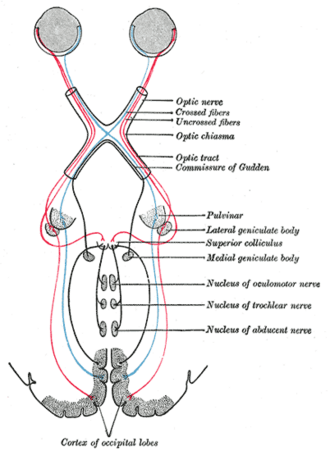
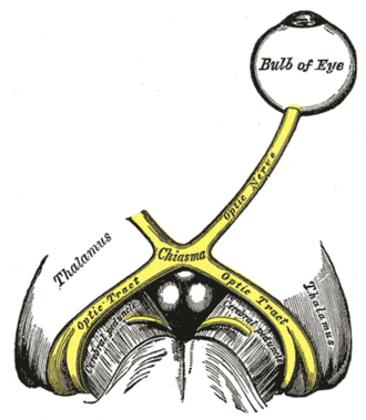
Optic nerve (orbital part, surrounded by meninges and CSF) → optic canal (with the ophthalmic artery) → optic chiasm above the pituitary, where fibers from the nasal half of each retina cross (carrying the temporal visual fields) → optic tract (ipsilateral temporal + contralateral nasal retina = the contralateral visual field) → lateral geniculate nucleus of the thalamus → optic radiations: the inferior fibers loop through the temporal lobe (Meyer loop) and carry the contralateral superior quadrant; the superior fibers pass through the parietal lobe and carry the inferior quadrant → primary visual cortex (V1) on the banks of the calcarine sulcus of the occipital lobe (see cerebral cortex and lobes).
- Branches
- Fibers from the optic tract to the pretectal nuclei (pupillary light reflex) and superior colliculus
- Retinohypothalamic fibers to the suprachiasmatic nucleus (circadian rhythm; see hypothalamus)
- Sensory
Vision (special somatic afferent); afferent limb of the pupillary light and accommodation reflexes.
- Relations
Inside the orbit it lies within the cone of the extraocular muscles; the central retinal artery enters it about 1 cm behind the eyeball. The chiasm sits above the pituitary gland and diaphragma sellae, with the internal carotid arteries lateral and the anterior cerebral/anterior communicating arteries above (see circle of Willis).
- Notes
Developmentally the optic nerve is a tract of the CNS — myelinated by oligodendrocytes and wrapped in meninges — which is why it is affected by MS and why raised intracranial pressure causes papilledema.
Found in
Mentioned in
Generated automatically from links in other entries.
Swipe sideways to see the whole diagram →


Clinical correlations
Visual field defects by lesion site PANCE
- Mechanism / lesion
Crossing of the nasal fibers at the chiasm and the split of the radiations between temporal and parietal lobes.
- Classic presentation
Optic nerve → monocular vision loss (ipsilateral). Chiasm (pituitary adenoma, craniopharyngioma) → bitemporal hemianopia. Optic tract → contralateral homonymous hemianopia (often incongruous). Meyer loop (temporal lobe) → contralateral superior quadrantanopia ("pie in the sky"). Parietal radiations → contralateral inferior quadrantanopia ("pie on the floor"). Occipital cortex (PCA stroke) → contralateral homonymous hemianopia with macular sparing (dual MCA/PCA supply of the occipital pole).
Optic neuritis PANCE
- Mechanism / lesion
Inflammatory demyelination — often the first presentation of multiple sclerosis; also neuromyelitis optica (aquaporin-4) and MOG antibody disease.
- Classic presentation
Subacute painful monocular vision loss, pain with eye movement, reduced color vision (red desaturation), a relative afferent pupillary defect, central scotoma; the disc is often normal (retrobulbar). MRI brain; IV methylprednisolone speeds recovery.
Papilledema PANCE
- Mechanism / lesion
Raised intracranial pressure transmitted along the optic nerve sheath (tumor, hemorrhage, venous sinus thrombosis, idiopathic intracranial hypertension).
- Classic presentation
Bilateral optic disc swelling with blurred margins, loss of venous pulsations, flame hemorrhages; transient visual obscurations, headache worse lying down, enlarged blind spot. Image before lumbar puncture.
Central retinal artery occlusion PANCE
- Mechanism / lesion
Embolus (carotid, cardiac) or giant cell arteritis blocking the central retinal artery.
- Classic presentation
Sudden painless monocular vision loss, RAPD, cherry-red spot with a pale, swollen retina. A stroke equivalent — urgent stroke workup; check ESR/CRP in older patients.
High-yield PANCE pearls
- Bitemporal hemianopia → chiasm (pituitary adenoma; craniopharyngioma in children).
- "Pie in the sky" → temporal lobe (Meyer loop); "pie on the floor" → parietal.
- Homonymous hemianopia with macular sparing → PCA stroke (occipital).
- Painful monocular vision loss + pain on eye movement + RAPD → optic neuritis (MS).
- Pupillary light reflex: afferent CN II, efferent CN III.
- Bilateral disc swelling = papilledema = raised ICP until proven otherwise.
Imaging anatomy
- CT
Emergency CT for trauma, hemorrhage and mass effect; CT venography when venous sinus thrombosis is suspected.
- MRI
MRI brain and orbits with contrast — optic neuritis (enhancing nerve, white matter lesions), pituitary and chiasmal lesions, tumors along the pathway.
- Key views & landmarks
- Formal perimetry (Humphrey visual fields) documents and follows defects
- Optical coherence tomography (OCT) of the retinal nerve fiber layer
Physical exam
- Surface landmarks
The optic disc on fundoscopy lies nasal to the macula; the physiologic blind spot lies about 15° temporal to fixation.
- Strength / function testing
Visual acuity (Snellen chart, each eye, with glasses or a pinhole); color vision (Ishihara plates); visual fields by confrontation (each eye, four quadrants); pupils — direct and consensual light reflex and accommodation; fundoscopy (disc margins, cup-to-disc ratio, venous pulsations, retina).
Swinging flashlight test
- Technique
Swing a bright light back and forth between the eyes, about 2–3 seconds on each, in a dim room.
- Positive test
Relative afferent pupillary defect (Marcus Gunn pupil) — both pupils dilate when the light moves to the affected eye.
- Significance
Asymmetric optic nerve (or extensive retinal) disease — optic neuritis, ischemic optic neuropathy, CRAO, severe glaucoma. Not caused by cataract or refractive error.
Self-quiz
A 45-year-old woman has headaches and keeps bumping into door frames on both sides. Confrontation testing shows loss of the temporal half of the visual field in each eye. Where is the lesion?
Correct answer: B
Bitemporal hemianopia results from compression of the crossing nasal fibers at the chiasm, classically by a pituitary macroadenoma. Next step: MRI of the sella and pituitary hormone testing.
Show answer
B
Bitemporal hemianopia results from compression of the crossing nasal fibers at the chiasm, classically by a pituitary macroadenoma. Next step: MRI of the sella and pituitary hormone testing.
A 28-year-old woman has 3 days of blurred vision in the right eye with pain on eye movement. When a light is swung from the left eye to the right, both pupils dilate. What is the most likely diagnosis?
Correct answer: B
Painful vision loss with pain on eye movement and a relative afferent pupillary defect in a young woman is optic neuritis, often the first sign of multiple sclerosis — obtain MRI.
Show answer
B
Painful vision loss with pain on eye movement and a relative afferent pupillary defect in a young woman is optic neuritis, often the first sign of multiple sclerosis — obtain MRI.
After a right temporal lobectomy, a patient has a visual field defect. Which defect is expected?
Correct answer: B
The Meyer loop in the temporal lobe carries fibers for the contralateral superior quadrant, so a right temporal lesion causes a left superior quadrantanopia ("pie in the sky").
Show answer
B
The Meyer loop in the temporal lobe carries fibers for the contralateral superior quadrant, so a right temporal lesion causes a left superior quadrantanopia ("pie in the sky").
A lesion of the optic radiations in the parietal lobe causes which visual field defect?
Correct answer: D
The parietal radiations carry the inferior field ("pie on the floor"); Meyer loop in the temporal lobe carries the superior field ("pie in the sky").
Show answer
D
The parietal radiations carry the inferior field ("pie on the floor"); Meyer loop in the temporal lobe carries the superior field ("pie in the sky").
Why is the optic nerve affected by demyelinating diseases such as multiple sclerosis?
Correct answer: A
Developmentally the optic nerve is a CNS tract, myelinated by oligodendrocytes and wrapped in meninges, which is why MS causes optic neuritis and raised intracranial pressure causes papilledema.
Show answer
A
Developmentally the optic nerve is a CNS tract, myelinated by oligodendrocytes and wrapped in meninges, which is why MS causes optic neuritis and raised intracranial pressure causes papilledema.