Spinal cord
Continuation of the medulla within the vertebral canal, ending at the conus medullaris at about L1–L2 in adults. Its blood supply and level of termination explain spinal cord injury syndromes and safe lumbar puncture sites.
Core anatomy
- Location
Vertebral canal, from the foramen magnum to the conus medullaris at about the L1–L2 disc in adults (lower, about L3, in newborns). verify
- Parts
- Cervical enlargement (about C4–T1, for the brachial plexus) and lumbosacral enlargement (about L1–S3 segments, for the lumbosacral plexus)
- Conus medullaris: tapered lower end; filum terminale continues to the coccyx
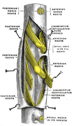
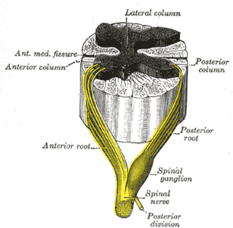
- 31 pairs of spinal nerves (8 C, 12 T, 5 L, 5 S, 1 Co) from dorsal (sensory) and ventral (motor) rootlets
- Internal organization: central gray matter (dorsal, lateral T1–L2 sympathetic, and ventral horns) surrounded by white matter tracts (dorsal columns, spinothalamic, corticospinal)
- Relations
Surrounded by the spinal meninges and spaces; roots below the conus form the cauda equina. Because the cord is shorter than the column, lower cord segments lie higher than their vertebrae (e.g. the lumbar segments lie at about T10–T12).
- Arterial supply
One anterior spinal artery (from the vertebral arteries) supplies the anterior two-thirds of the cord. Two posterior spinal arteries supply the posterior third (dorsal columns). They are reinforced by segmental medullary arteries, the largest being the great anterior segmental medullary artery (of Adamkiewicz), usually on the left between about T9 and T12, supplying the lower cord. verify
- Venous drainage
Spinal veins → internal vertebral venous plexus (vertebral venous plexus).
Mentioned in
- Accessory nerve (CN XI)
- Dorsal column–medial lemniscus pathway
- Medulla oblongata
- Spinal meninges and spaces
- Spinothalamic tract
- Vertebral column
Generated automatically from links in other entries.
Swipe sideways to see the whole diagram →


Clinical correlations
Anterior spinal artery (anterior cord) syndrome PANCE
- Mechanism / lesion
Infarction of the anterior two-thirds of the cord, e.g. after aortic surgery or dissection (loss of the artery of Adamkiewicz), hypotension, or flexion injury.
- Classic presentation
Paraplegia or quadriplegia and loss of pain and temperature below the lesion, with preserved proprioception and vibration (dorsal columns). Bladder dysfunction.
Incomplete cord injury syndromes PANCE
- Mechanism / lesion
Central cord: hyperextension in an older patient with cervical spondylosis. Brown-Séquard: hemisection (penetrating trauma). See also the anterior cord syndrome above.
- Classic presentation
Central cord: weakness greater in the arms than the legs, variable sensory loss. Brown-Séquard: ipsilateral weakness and loss of proprioception/vibration, contralateral loss of pain and temperature below the lesion.
Neurogenic shock PANCE
- Mechanism / lesion
Loss of sympathetic outflow after cervical or high thoracic (above about T6) cord injury.
- Classic presentation
Hypotension with bradycardia and warm, dry skin (unlike hemorrhagic shock with tachycardia). Treat with fluids and vasopressors.
Neural tube defects PANCE
- Mechanism / lesion
Failure of neural tube closure. Risk reduced by periconceptional folic acid; elevated maternal AFP.
- Classic presentation
Spina bifida occulta: bony defect with a tuft of hair or dimple, usually asymptomatic. Meningocele: meninges protrude. Myelomeningocele: cord and meninges protrude, with leg paralysis, bladder dysfunction, and Chiari II malformation/hydrocephalus. Tethered cord: low-lying conus causing back pain, gait and bladder problems.
High-yield PANCE pearls
- Cord ends at L1–L2 in adults → LP at L3–L4 or L4–L5.
- Anterior spinal artery syndrome: motor + pain/temperature loss, preserved vibration/proprioception; after aortic surgery (Adamkiewicz).
- Central cord: arms > legs, older patient, hyperextension. Brown-Séquard: ipsilateral motor/dorsal columns, contralateral pain/temperature.
- Neurogenic shock: hypotension + bradycardia after high cord injury.
- Folic acid prevents neural tube defects; hair tuft over the spine → spina bifida occulta.
Imaging anatomy
- X-ray
Plain films show bony injury and alignment only.
- CT
Bony injury in trauma; CT myelography when MRI is contraindicated.
- MRI
MRI is the test of choice for the cord: compression, contusion/edema, infarction (diffusion changes), demyelination, syrinx, tumor, tethered cord.
- Ultrasound
In infants (before posterior element ossification), spinal ultrasound evaluates a low-lying conus and tethered cord (e.g. a sacral dimple).
- Key views & landmarks
- MRI spine (sagittal T2 and STIR, axial T2)
Physical exam
- Surface landmarks
The conus lies at about the L1–L2 vertebral level; the intercristal line (L4) is safely below it.
- Strength / function testing
Localize by sensory level, myotomes and reflexes; UMN signs below the lesion (spasticity, hyperreflexia, Babinski) and LMN signs at the level. Assess rectal tone and perianal sensation (sacral sparing indicates an incomplete injury). The ASIA/ISNCSCI exam grades injury severity.
Self-quiz
After repair of a thoracoabdominal aortic aneurysm, a patient cannot move his legs and cannot feel pinprick below the umbilicus, but vibration and position sense are preserved. Which vessel was compromised?
Correct answer: B
Anterior spinal artery syndrome spares the dorsal columns (vibration, proprioception). The lower cord's anterior supply depends on the artery of Adamkiewicz, which can be lost during aortic surgery.
Show answer
B
Anterior spinal artery syndrome spares the dorsal columns (vibration, proprioception). The lower cord's anterior supply depends on the artery of Adamkiewicz, which can be lost during aortic surgery.
A 70-year-old man falls forward and hits his chin, hyperextending his neck. He has marked weakness of both hands with much milder leg weakness. What is the most likely syndrome?
Correct answer: B
Central cord syndrome (arms weaker than legs) typically follows hyperextension in an older patient with cervical spondylosis.
Show answer
B
Central cord syndrome (arms weaker than legs) typically follows hyperextension in an older patient with cervical spondylosis.
A patient with a C5 spinal cord injury is hypotensive (80/40) with a heart rate of 48 and warm extremities. What is the cause?
Correct answer: B
Loss of sympathetic tone after high cord injury causes hypotension with bradycardia and warm skin: neurogenic shock.
Show answer
B
Loss of sympathetic tone after high cord injury causes hypotension with bradycardia and warm skin: neurogenic shock.
At what vertebral level does the spinal cord typically end in adults?
Correct answer: C
The conus medullaris ends at about L1–L2 in adults, so lumbar puncture is performed at L3–L4 or L4–L5.
Show answer
C
The conus medullaris ends at about L1–L2 in adults, so lumbar puncture is performed at L3–L4 or L4–L5.
After a stab wound to the right side of the thoracic spine, which pattern of deficits is expected below the lesion?
Correct answer: D
Brown-Séquard (hemisection) causes ipsilateral weakness (corticospinal) and loss of vibration/proprioception (dorsal columns), with contralateral loss of pain and temperature because the spinothalamic fibers have already crossed.
Show answer
D
Brown-Séquard (hemisection) causes ipsilateral weakness (corticospinal) and loss of vibration/proprioception (dorsal columns), with contralateral loss of pain and temperature because the spinothalamic fibers have already crossed.