Deltoid
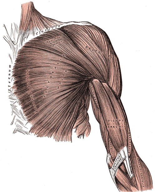
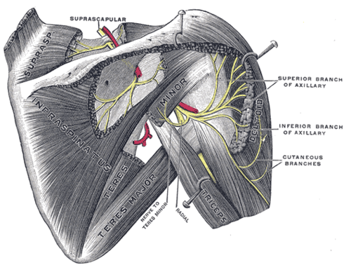
Triangular muscle that forms the rounded contour of the shoulder. It is the main abductor of the arm beyond the first ~15° and is supplied by the axillary nerve.
Core anatomy
- Location
Covers the glenohumeral joint anteriorly, laterally and posteriorly, forming the shoulder contour.
- Group
Superficial scapulohumeral muscles; three parts (clavicular/anterior, acromial/middle, spinal/posterior).
- Origin
Lateral third of the clavicle (anterior part), acromion (middle part), and spine of the scapula (posterior part).
- Insertion
Deltoid tuberosity of the humerus.
- Innervation
Axillary nerve (C5, C6)
- Blood supply
Posterior circumflex humeral artery (principal) and the deltoid branch of the thoracoacromial artery verify
- Action
- Anterior (clavicular) part: flexes and medially rotates the arm
- Middle (acromial) part: abducts the arm after supraspinatus initiates the movement
- Posterior (spinal) part: extends and laterally rotates the arm
- Relations
The axillary nerve and posterior circumflex humeral artery run on its deep surface around the surgical neck of the humerus. The subacromial–subdeltoid bursa lies between deltoid and the rotator cuff. The deltopectoral groove (with the cephalic vein) separates its anterior border from pectoralis major.


Clinical correlations
Deltoid paralysis (axillary nerve injury) PANCE
- Mechanism / lesion
Surgical neck fracture of the humerus, anterior shoulder dislocation, or iatrogenic injury.
- Classic presentation
Weak abduction beyond the initial range. Over weeks the muscle atrophies, flattening the rounded contour of the shoulder. Numbness over the regimental badge area.
Intramuscular injection site
- Mechanism / lesion
Deltoid is a common IM injection site. The axillary nerve runs horizontally on its deep surface around the surgical neck, so injections too low or too posterior risk the nerve. Injections placed too high risk the subacromial bursa (shoulder injury related to vaccine administration, SIRVA).
- Classic presentation
Inject into the thickest part of the middle deltoid, about 2–3 fingerbreadths (≈5 cm) below the acromion. verify
High-yield PANCE pearls
- Deltoid atrophy + lateral shoulder numbness → axillary nerve (surgical neck fracture, anterior dislocation).
- Supraspinatus initiates abduction (~0–15°); deltoid continues it.
- A 'squared-off' shoulder in acute anterior dislocation comes from the displaced humeral head, not deltoid paralysis.
Imaging anatomy
- X-ray
Soft-tissue contour only. Look at the humerus for surgical neck fractures that endanger its nerve supply.
- MRI
Denervation edema (acute) or fatty atrophy (chronic) with axillary neuropathy. Rarely, deltoid tears or avulsions.
- Ultrasound
Superficial muscle over the cuff. Ultrasound shows deltoid thickness when guiding IM injections and visualizes the subdeltoid bursa beneath it.
Physical exam
- Surface landmarks
Rounded contour of the shoulder. The deltoid tuberosity is palpable roughly mid-way down the lateral arm.
- Palpation
Palpate all three parts. Compare bulk with the other side and look for hollowing below the acromion.
- Strength / function testing
Resisted abduction with the arm at about 90° (middle part), resisted flexion (anterior part), and resisted extension (posterior part). Check sensation over the lateral shoulder.
Self-quiz
Three months after a proximal humerus fracture, a 70-year-old woman has flattening of the lateral shoulder contour and can't abduct her arm beyond about 15°. Which nerve was injured?
Correct answer: B
The axillary nerve supplies deltoid and wraps around the surgical neck. Supraspinatus (suprascapular nerve) still initiates abduction, but the patient can't continue it without deltoid, and the muscle atrophies.
Show answer
B
The axillary nerve supplies deltoid and wraps around the surgical neck. Supraspinatus (suprascapular nerve) still initiates abduction, but the patient can't continue it without deltoid, and the muscle atrophies.
Which part of the deltoid laterally rotates the arm?
Correct answer: C
The spinal (posterior) part extends and laterally rotates. The clavicular (anterior) part flexes and medially rotates. The acromial (middle) part abducts.
Show answer
C
The spinal (posterior) part extends and laterally rotates. The clavicular (anterior) part flexes and medially rotates. The acromial (middle) part abducts.
The deltoid inserts on which structure?
Correct answer: C
All three parts of deltoid converge on the deltoid tuberosity on the lateral surface of the humeral shaft.
Show answer
C
All three parts of deltoid converge on the deltoid tuberosity on the lateral surface of the humeral shaft.
A patient with an acute anterior shoulder dislocation has a "squared-off" shoulder. What causes this contour?
Correct answer: B
The squared-off shoulder of an acute anterior dislocation comes from the displaced humeral head (moved anteroinferiorly), leaving the acromion prominent. It is not deltoid paralysis. Flattening from deltoid atrophy after axillary nerve injury develops over weeks.
Show answer
B
The squared-off shoulder of an acute anterior dislocation comes from the displaced humeral head (moved anteroinferiorly), leaving the acromion prominent. It is not deltoid paralysis. Flattening from deltoid atrophy after axillary nerve injury develops over weeks.
Which vein runs in the groove between deltoid and pectoralis major?
Correct answer: C
The cephalic vein runs in the deltopectoral groove between the anterior border of deltoid and pectoralis major before draining into the axillary vein.
Show answer
C
The cephalic vein runs in the deltopectoral groove between the anterior border of deltoid and pectoralis major before draining into the axillary vein.
Flashcards
See also
- Axillary nerve
- Supraspinatus
- A shoulder forced back while blocking (clinical case)