Elbow joint
Hinge joint between the humerus, ulna and radius. It shares a capsule with the proximal radioulnar joint and is the site of common overuse and pediatric injuries.
Core anatomy
- Location
Between the distal humerus and the proximal ulna and radius.
- Classification
Synovial hinge joint (humeroulnar and humeroradial articulations). The proximal radioulnar joint (a pivot joint) lies within the same capsule.
- Articulating surfaces
Trochlea of the humerus with the trochlear notch of the ulna; capitulum of the humerus with the head of the radius.
- Capsule
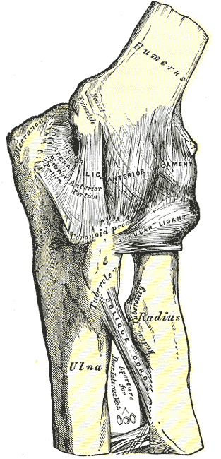
Fibrous capsule, thin anteriorly and posteriorly, reinforced on each side by the collateral ligaments.
- Ligaments
- Ulnar (medial) collateral ligament: medial epicondyle to the coronoid process and olecranon; anterior, posterior and oblique bands. The anterior band is the strongest and the main restraint to valgus stress
- Radial (lateral) collateral ligament: lateral epicondyle, blending with the anular ligament; resists varus stress
- Anular ligament: encircles and holds the radial head in the radial notch of the ulna
- Movements
- Flexion and extension (humeroulnar and humeroradial)
- Pronation and supination occur at the proximal and distal radioulnar joints
- Normal carrying angle (cubitus valgus) of about 5–15°, typically greater in females verify
- Stability
Bony congruity of the trochlea and trochlear notch, plus the collateral ligaments and surrounding muscles.
- Innervation
Musculocutaneous nerve, radial nerve, ulnar nerve and median nerve (articular branches)
- Blood supply
Periarticular arterial anastomosis (collateral branches of the brachial and profunda brachii; recurrent branches of the radial, ulnar and interosseous arteries).
- Relations
Anteriorly the cubital fossa (brachial artery, median nerve, biceps tendon). Posteriorly the ulnar nerve lies behind the medial epicondyle, and the olecranon bursa is subcutaneous over the olecranon.

Clinical correlations
Lateral epicondylitis (tennis elbow) PANCE
- Mechanism / lesion
Degenerative tendinopathy (tendinosis) of the common extensor origin, particularly extensor carpi radialis brevis (ECRB), from repetitive wrist extension and gripping.
- Classic presentation
Lateral elbow pain and tenderness just distal to the lateral epicondyle. Pain with resisted wrist extension (Cozen test) and with passive wrist flexion with the elbow extended (Mill test). Grip weakness from pain.
Medial epicondylitis (golfer's elbow) PANCE
- Mechanism / lesion
Tendinopathy of the common flexor–pronator origin (pronator teres and FCR) from repetitive wrist flexion and pronation.
- Classic presentation
Medial epicondyle pain worsened by resisted wrist flexion and pronation. May coexist with ulnar neuropathy.
Elbow dislocation PANCE
- Mechanism / lesion
Fall on an outstretched hand. Posterior dislocation is most common.
- Classic presentation
Deformity with a prominent olecranon (the epicondyle–olecranon triangle is disrupted). Check the brachial artery, median nerve and ulnar nerve. Associated fractures (radial head, coronoid) can form the "terrible triad" (dislocation + radial head fracture + coronoid fracture).
Ulnar collateral ligament injury
- Mechanism / lesion
Repetitive valgus stress in overhead throwing athletes (pitchers, javelin), or acute rupture.
- Classic presentation
Medial elbow pain during the late cocking/early acceleration phase of throwing, reduced velocity, and ulnar nerve symptoms. Valgus stress testing is painful or lax.
High-yield PANCE pearls
- Lateral epicondylitis = ECRB; pain with resisted wrist extension (Cozen).
- Medial epicondylitis = flexor–pronator origin; pain with resisted wrist flexion and pronation.
- Posterior fat pad sign = effusion = occult fracture until proven otherwise.
- Pediatric elbow ossification order: CRITOE: Capitellum, Radial head, Internal (medial) epicondyle, Trochlea, Olecranon, External (lateral) epicondyle.
- Posterior dislocation is the most common elbow dislocation.
Imaging anatomy
- X-ray
AP and true lateral with the elbow at 90°. Look for: fat pads (a visible posterior fat pad or elevated anterior "sail" fat pad = effusion); the anterior humeral line (through the middle third of the capitellum); the radiocapitellar line (through the capitellum in all views). In children, interpret ossification centers using CRITOE.
- CT
Complex fractures (terrible triad, distal humerus intra-articular fractures).
- MRI
UCL and LCL tears, epicondylitis (common tendon thickening and increased signal), osteochondral lesions (e.g. osteochondritis dissecans of the capitellum in young throwers).
- Ultrasound
Common extensor/flexor tendinopathy (hypoechoic thickening, neovascularity on Doppler), UCL assessment with dynamic valgus stress, effusion, olecranon bursitis, and ulnar nerve subluxation.
- Key views & landmarks
- Elbow AP and lateral at 90°
- Radial head–capitellum view (oblique) for subtle radial head fractures
Physical exam
- Surface landmarks
Medial and lateral epicondyles, olecranon (straight line in extension, isosceles triangle in 90° flexion), radial head (distal to the lateral epicondyle, rotates with pronation/supination), ulnar nerve in the groove behind the medial epicondyle.
- Palpation
Palpate both epicondyles, the radial head, the olecranon and bursa, and the distal biceps tendon.
- Strength / function testing
Flexion, extension, pronation and supination (active and passive). Varus and valgus stress at about 20–30° of flexion.
Cozen test
- Technique
Elbow extended (or slightly flexed), forearm pronated. The patient makes a fist and extends and radially deviates the wrist against resistance.
- Positive test
Pain at the lateral epicondyle.
- Significance
Lateral epicondylitis.
Mill test
- Technique
With the elbow extended and forearm pronated, the examiner passively flexes the wrist fully.
- Positive test
Pain at the lateral epicondyle.
- Significance
Lateral epicondylitis.
Golfer's elbow test
- Technique
Elbow extended. The patient flexes the wrist and pronates the forearm against resistance.
- Positive test
Pain at the medial epicondyle.
- Significance
Medial epicondylitis.
Valgus stress test
- Technique
Elbow flexed about 20–30° to unlock the olecranon. The examiner applies a valgus force.
- Positive test
Medial pain and/or increased laxity compared with the other side.
- Significance
UCL sprain or tear.
Self-quiz
A 45-year-old carpenter has lateral elbow pain for 2 months. Pain is reproduced by resisted wrist extension with the elbow extended and forearm pronated. Which tendon is most commonly involved?
Correct answer: B
Lateral epicondylitis is a tendinopathy of the common extensor origin, most characteristically ECRB. Pronator teres and FCR are involved in medial epicondylitis.
Show answer
B
Lateral epicondylitis is a tendinopathy of the common extensor origin, most characteristically ECRB. Pronator teres and FCR are involved in medial epicondylitis.
A lateral radiograph of a child's elbow shows an elevated anterior fat pad and a visible posterior fat pad. What does this indicate?
Correct answer: B
A visible posterior fat pad is never normal. It indicates an effusion, which after trauma suggests an occult fracture (supracondylar in children, radial head in adults). Nursemaid's elbow has normal radiographs.
Show answer
B
A visible posterior fat pad is never normal. It indicates an effusion, which after trauma suggests an occult fracture (supracondylar in children, radial head in adults). Nursemaid's elbow has normal radiographs.
Which ligament is the primary restraint to valgus stress at the elbow in an overhead throwing athlete?
Correct answer: C
The anterior band of the UCL is the key restraint to valgus stress and is injured by repetitive throwing.
Show answer
C
The anterior band of the UCL is the key restraint to valgus stress and is injured by repetitive throwing.
The "terrible triad" of the elbow consists of which injuries?
Correct answer: D
The terrible triad is an elbow dislocation with fractures of the radial head and the coronoid process. Posterior dislocation is the most common elbow dislocation; check the brachial artery, median nerve and ulnar nerve.
Show answer
D
The terrible triad is an elbow dislocation with fractures of the radial head and the coronoid process. Posterior dislocation is the most common elbow dislocation; check the brachial artery, median nerve and ulnar nerve.
Using the mnemonic CRITOE, which ossification center of the pediatric elbow appears last?
Correct answer: E
CRITOE: Capitellum, Radial head, Internal (medial) epicondyle, Trochlea, Olecranon, External (lateral) epicondyle. Knowing the order helps tell a normal unfused center from a fracture fragment on a child's elbow radiograph.
Show answer
E
CRITOE: Capitellum, Radial head, Internal (medial) epicondyle, Trochlea, Olecranon, External (lateral) epicondyle. Knowing the order helps tell a normal unfused center from a fracture fragment on a child's elbow radiograph.