Abdominal aorta
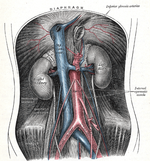
Continuation of the thoracic aorta from the aortic hiatus (T12) to its bifurcation at L4. It supplies the abdominal organs and is the usual site of aortic aneurysm.
Core anatomy
- Location
Retroperitoneal, on the anterior surface of the lumbar vertebral bodies, slightly left of the midline, with the inferior vena cava on its right.
- Origin
Continuation of the descending thoracic aorta at the aortic hiatus of the diaphragm (T12).
- Course
Descends anterior to the lumbar vertebrae to bifurcate at about L4 (roughly at or slightly below the umbilicus) into the right and left common iliac arteries.
- Branches
- Anterior (unpaired, to the gut): Celiac trunk (T12), superior mesenteric artery (L1), inferior mesenteric artery (L3)
- Lateral (paired): inferior phrenic, middle suprarenal, renal (about L1–L2), gonadal (about L2)
- Posterior: lumbar arteries (four pairs), median sacral artery
- Terminal: right and left common iliac arteries (L4)
- Supplies
Abdominal viscera, posterior abdominal wall, kidneys, suprarenal glands, gonads, and (via the iliacs) the pelvis and lower limbs.
- Termination
Common iliac arteries at L4.
- Relations
Anterior: pancreas and splenic vein, the left renal vein (which crosses between the aorta and the SMA), the 3rd part of the duodenum, and the root of the mesentery. Right: IVC, cisterna chyli, azygos vein. The sympathetic trunks run on each side of the vertebral bodies.
Branches
Structures supplied
Mentioned in
Generated automatically from links in other entries.
Swipe sideways to see the whole diagram →

Clinical correlations
Abdominal aortic aneurysm (AAA) PANCE
- Mechanism / lesion
Dilation of the aorta to 3.0 cm or more, usually infrarenal. Risk factors: smoking (the strongest), male sex, age over 65, family history, hypertension, atherosclerosis.
- Classic presentation
Usually asymptomatic, found on screening or incidental imaging. Rupture presents with the triad of severe abdominal or back/flank pain, hypotension and a pulsatile abdominal mass. Elective repair is generally recommended at 5.5 cm or more in men and 5.0 cm or more in women, with rapid growth, or when symptomatic. Surveillance ultrasound (SVS 2018): every 3 years at 3.0–3.9 cm, every 12 months at 4.0–4.9 cm and every 6 months at 5.0–5.4 cm.
AAA screening PANCE
- Mechanism / lesion
Screening detects asymptomatic aneurysms before rupture.
- Classic presentation
USPSTF: one-time abdominal ultrasound for men aged 65–75 who have ever smoked.
Aortoiliac occlusive disease (Leriche syndrome)
- Mechanism / lesion
Atherosclerotic occlusion of the distal aorta and/or iliac arteries.
- Classic presentation
Classic triad of buttock/thigh claudication, erectile dysfunction, and absent or diminished femoral pulses.
High-yield PANCE pearls
- AAA = ≥ 3.0 cm; most are infrarenal; smoking is the strongest risk factor.
- Screen: one-time ultrasound, men 65–75 who ever smoked (USPSTF).
- Rupture triad: pain + hypotension + pulsatile mass → emergency surgery (unstable patients go straight to the OR).
- Leriche: buttock claudication + ED + absent femoral pulses.
- Branch levels: celiac T12, SMA L1, renals L1–L2, IMA L3, bifurcation L4.
Imaging anatomy
- X-ray
Calcified aneurysm wall ("eggshell" calcification) on a lateral abdominal film. Not reliable for sizing.
- CT
CT angiography is the test of choice for stable patients with suspected rupture and for planning repair (open or endovascular). It measures diameter, the neck length below the renal arteries, and iliac involvement.
- Ultrasound
Screening and surveillance test (outer-to-outer wall diameter). Point-of-care ultrasound in the ED can quickly confirm an aneurysm in an unstable patient (though it cannot reliably show retroperitoneal rupture).
- Key views & landmarks
- Abdominal ultrasound (transverse and longitudinal aorta)
- CT angiography abdomen/pelvis
Physical exam
- Surface landmarks
The bifurcation lies at about L4, projecting near the level of the iliac crests (supracristal plane), slightly below the umbilicus.
- Palpation
Palpate deeply above and to the left of the umbilicus with both hands to estimate the width of the aortic pulsation (an expansile pulsation wider than about 3 cm suggests AAA). Sensitivity is limited by body habitus. Don't palpate forcefully if a symptomatic aneurysm is suspected.
- Strength / function testing
Auscultate for bruits (aortic, renal, iliac). Check femoral and distal pulses.
Self-quiz
A 72-year-old man with a 50-pack-year smoking history has sudden severe back pain, BP 76/40, and a tender pulsatile periumbilical mass. What is the next step?
Correct answer: B
The rupture triad (pain, hypotension, pulsatile mass) in an unstable patient calls for immediate repair. CT angiography is only for hemodynamically stable patients.
Show answer
B
The rupture triad (pain, hypotension, pulsatile mass) in an unstable patient calls for immediate repair. CT angiography is only for hemodynamically stable patients.
Which patient should be screened for abdominal aortic aneurysm under USPSTF guidance?
Correct answer: B
USPSTF recommends a one-time ultrasound for men aged 65–75 who have ever smoked. Screening in women who have never smoked isn't recommended.
Show answer
B
USPSTF recommends a one-time ultrasound for men aged 65–75 who have ever smoked. Screening in women who have never smoked isn't recommended.
At approximately which vertebral level does the abdominal aorta bifurcate?
Correct answer: D
The aorta bifurcates into the common iliac arteries at about L4. The IVC forms at about L5.
Show answer
D
The aorta bifurcates into the common iliac arteries at about L4. The IVC forms at about L5.
A 58-year-old smoker has cramping pain in both buttocks and thighs when walking, erectile dysfunction, and diminished femoral pulses. What is the most likely diagnosis?
Correct answer: D
Leriche syndrome (atherosclerotic occlusion of the distal aorta and/or iliac arteries) causes the triad of buttock/thigh claudication, erectile dysfunction and absent or diminished femoral pulses. Superficial femoral disease causes calf claudication with normal femoral pulses.
Show answer
D
Leriche syndrome (atherosclerotic occlusion of the distal aorta and/or iliac arteries) causes the triad of buttock/thigh claudication, erectile dysfunction and absent or diminished femoral pulses. Superficial femoral disease causes calf claudication with normal femoral pulses.
Which vessel crosses anterior to the abdominal aorta, just below the origin of the superior mesenteric artery?
Correct answer: A
The left renal vein crosses anterior to the aorta, between it and the SMA. Compression there ("nutcracker") can cause hematuria and a left varicocele. The right renal artery passes behind the IVC.
Show answer
A
The left renal vein crosses anterior to the aorta, between it and the SMA. Compression there ("nutcracker") can cause hematuria and a left varicocele. The right renal artery passes behind the IVC.