Spermatic cord
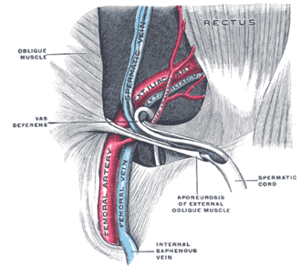
Bundle of structures running between the deep inguinal ring and the testis. It carries the ductus deferens, testicular vessels and pampiniform plexus, and is involved in varicocele, torsion and inguinal hernia.
Core anatomy
- Location
From the deep inguinal ring, through the inguinal canal, to the posterior border of the testis in the scrotum.
- Parts
- Ductus (vas) deferens and the artery of the ductus deferens
- Testicular artery (from the abdominal aorta at about L2)
- Pampiniform venous plexus → testicular vein
- Cremasteric artery and vein
- Genital branch of the genitofemoral nerve (supplies cremaster)
- Sympathetic and visceral afferent fibers; lymphatics (drain to lumbar/para-aortic nodes)
- Remnant of the processus vaginalis
- Coverings (derived from abdominal wall layers): external spermatic fascia (external oblique), cremasteric muscle and fascia (internal oblique), internal spermatic fascia (transversalis fascia)
- Relations
An indirect inguinal hernia sac lies within the coverings of the cord, anterolateral to its contents. The ilioinguinal nerve runs on the cord but outside its coverings.
- Arterial supply
Testicular artery, artery of the ductus deferens (from the inferior vesical/umbilical artery), cremasteric artery (from the inferior epigastric artery).
- Venous drainage
Pampiniform plexus → testicular vein. The right testicular vein drains into the IVC at an oblique angle; the left drains into the left renal vein at a right angle.
- Lymphatic drainage
Testicular lymph drains to the lumbar (para-aortic) nodes, not the inguinal nodes. Scrotal skin drains to the superficial inguinal nodes.
- Innervation
Genital branch of the genitofemoral nerve (L1–L2) to cremaster; testicular plexus (visceral, about T10).
Found in
Mentioned in
Generated automatically from links in other entries.

Clinical correlations
Varicocele PANCE
- Mechanism / lesion
Dilation of the pampiniform plexus, about 90% on the left (the left testicular vein drains at a right angle into the left renal vein, which can be compressed between the aorta and SMA). verify
- Classic presentation
A "bag of worms" above the testis that is more prominent standing or with Valsalva and decreases lying down. Associated with infertility and testicular atrophy. A new right-sided or non-decompressing varicocele (doesn't shrink supine) warrants imaging for a retroperitoneal mass (e.g. renal cell carcinoma) or IVC obstruction.
Testicular torsion PANCE
- Mechanism / lesion
Twisting of the cord, usually in adolescents with a "bell clapper" deformity (the testis lacks normal posterior fixation), cutting off testicular blood flow.
- Classic presentation
Sudden severe scrotal pain, nausea/vomiting, a high-riding, horizontally oriented testis, and an absent cremasteric reflex. A surgical emergency: salvage rates fall sharply after about 6 hours. Don't delay surgery for imaging when clinical suspicion is high.
Hydrocele PANCE
- Mechanism / lesion
Fluid in the tunica vaginalis (communicating, via a patent processus vaginalis, or non-communicating).
- Classic presentation
Painless scrotal swelling that transilluminates. Communicating hydroceles change size during the day.
High-yield PANCE pearls
- Varicocele: left > right, bag of worms; right-sided or non-reducing → image for RCC/IVC obstruction.
- Torsion: sudden pain, high horizontal testis, absent cremasteric reflex → immediate urology/surgery.
- Cremasteric reflex = L1–L2 (genitofemoral nerve).
- Testicular cancer spreads to para-aortic nodes (not inguinal).
- Cord coverings: external spermatic (EO), cremasteric (IO), internal spermatic (transversalis fascia).
Imaging anatomy
- CT
Evaluates the retroperitoneum when a varicocele is right-sided or doesn't decompress supine; staging of testicular cancer (para-aortic nodes).
- Ultrasound
Scrotal ultrasound with color Doppler is first-line: absent or reduced flow and a twisted cord ("whirlpool sign") in torsion; dilated veins (roughly 3 mm or more) with reflux on Valsalva in varicocele; an anechoic collection around the testis in hydrocele. verify
- Key views & landmarks
- Scrotal ultrasound with color Doppler, supine and with Valsalva/standing
Physical exam
- Surface landmarks
The cord is palpable between the finger and thumb at the neck of the scrotum, with the ductus deferens as a firm cord within it.
- Palpation
Palpate the cord standing and supine, with and without Valsalva (varicocele). Transilluminate scrotal swellings.
- Strength / function testing
Cremasteric reflex: stroke the superomedial thigh; the ipsilateral testis should rise. Absence suggests torsion. Testicular lie and position (high-riding, horizontal).
Self-quiz
A 14-year-old boy wakes with sudden severe left scrotal pain and vomiting. The left testis is high-riding with a horizontal lie, and the left cremasteric reflex is absent. What is the next step?
Correct answer: B
These findings indicate testicular torsion, a surgical emergency. When clinical suspicion is high, proceed to surgical exploration without delaying for imaging; testicular salvage declines rapidly after about 6 hours.
Show answer
B
These findings indicate testicular torsion, a surgical emergency. When clinical suspicion is high, proceed to surgical exploration without delaying for imaging; testicular salvage declines rapidly after about 6 hours.
A 58-year-old man has a new right-sided varicocele that does not decrease when he lies supine. What is the most important next step?
Correct answer: C
The right testicular vein drains directly into the IVC, so a new right-sided or non-decompressing varicocele raises concern for IVC or renal vein obstruction (e.g. renal cell carcinoma). Image the abdomen.
Show answer
C
The right testicular vein drains directly into the IVC, so a new right-sided or non-decompressing varicocele raises concern for IVC or renal vein obstruction (e.g. renal cell carcinoma). Image the abdomen.
The cremasteric muscle and fascia of the spermatic cord are derived from which layer of the abdominal wall?
Correct answer: B
Internal oblique → cremasteric muscle and fascia. External oblique → external spermatic fascia. Transversalis fascia → internal spermatic fascia.
Show answer
B
Internal oblique → cremasteric muscle and fascia. External oblique → external spermatic fascia. Transversalis fascia → internal spermatic fascia.
A painless scrotal swelling transilluminates brightly. What is the most likely diagnosis?
Correct answer: C
A hydrocele is fluid in the tunica vaginalis and transilluminates. A varicocele feels like a "bag of worms"; testicular tumors are solid and don't transilluminate.
Show answer
C
A hydrocele is fluid in the tunica vaginalis and transilluminates. A varicocele feels like a "bag of worms"; testicular tumors are solid and don't transilluminate.
A testicular cancer first spreads to which lymph nodes?
Correct answer: E
The testis develops on the posterior abdominal wall and descends with its vessels, so testicular lymph drains to the lumbar (para-aortic) nodes. Scrotal skin drains to the superficial inguinal nodes.
Show answer
E
The testis develops on the posterior abdominal wall and descends with its vessels, so testicular lymph drains to the lumbar (para-aortic) nodes. Scrotal skin drains to the superficial inguinal nodes.
Flashcards
See also
- Inguinal canal
- Abdominal aorta
- Kidney
- Sudden scrotal pain at night (clinical case)