Perineum
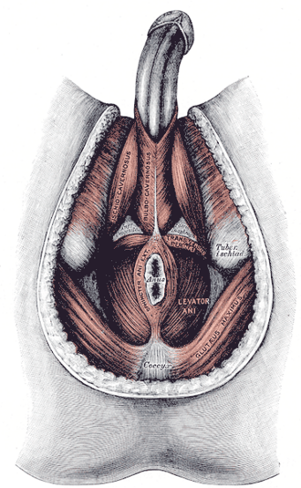
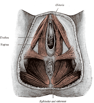
Diamond-shaped region below the pelvic diaphragm, divided into the urogenital and anal triangles. The perineal body at its center is torn in obstetric lacerations, and saddle anesthesia here signals cauda equina syndrome.
Core anatomy
- Location
Between the thighs, below the pelvic diaphragm and within the pelvic outlet.
- Boundaries
- anterior
- Inferior margin of the pubic symphysis
- anterolateral
- Ischiopubic rami
- lateral
- Ischial tuberosities
- posterolateral
- Sacrotuberous ligaments
- posterior
- Tip of the coccyx
- roof
- Pelvic diaphragm (levator ani and coccygeus)
- floor
- Skin and superficial fascia
- Contents
- Urogenital triangle (anterior, in front of a line between the ischial tuberosities; tilted forward): the perineal membrane, perineal pouches, external genitalia (penis and scrotum or vulva), and the urethra (and vagina)
- Anal triangle (posterior; tilted backward): the anal canal with the external anal sphincter, the two ischioanal fossae and the anococcygeal body
- Perineal body (central tendon of the perineum): a fibromuscular mass at the midpoint between the anus and the bulb of the penis or the vaginal vestibule, where bulbospongiosus, the external anal sphincter, the superficial and deep transverse perineal muscles and fibers of levator ani converge
- Nerves and vessels: branches of the pudendal nerve and internal pudendal vessels
- Communications
The superficial perineal pouch opens anteriorly into the space deep to the membranous layer of the anterior abdominal wall (Scarpa fascia). The ischioanal fossae extend forward above the perineal membrane as anterior recesses and communicate with each other behind the anal canal.
Mentioned in
Generated automatically from links in other entries.
Swipe sideways to see the whole diagram →


Clinical correlations
Obstetric perineal lacerations PANCE
- Mechanism / lesion
Stretching and tearing of the perineum during vaginal delivery; risk increases with operative delivery, a large baby, first delivery and midline episiotomy.
- Classic presentation
1st degree: perineal skin or vaginal epithelium only. 2nd degree: perineal muscles (perineal body) but not the anal sphincter. 3rd degree: the anal sphincter complex (3a < 50% of the external sphincter; 3b > 50%; 3c internal sphincter also torn). 4th degree: through the sphincters into the anorectal mucosa. Missed sphincter injuries cause later fecal incontinence.
- Notes
A mediolateral episiotomy is less likely than a midline one to extend into the sphincter; routine episiotomy is not recommended.
Saddle anesthesia (cauda equina syndrome) PANCE
- Mechanism / lesion
Compression of the S2–S5 roots in the cauda equina (central disc herniation, tumor, epidural abscess or hematoma).
- Classic presentation
Numbness of the perineum, buttocks and inner thighs, urinary retention with overflow, fecal incontinence, reduced anal tone, and sciatica. Emergency MRI and surgical decompression.
Fournier gangrene PANCE
- Mechanism / lesion
Polymicrobial necrotizing fasciitis of the perineum and genitalia, spreading along fascial planes (Colles, dartos, Scarpa). Risk factors — diabetes, obesity, alcohol use, immunosuppression, perianal or urinary infection, and SGLT2 inhibitors.
- Classic presentation
Severe perineal/genital pain out of proportion to the exam, swelling, erythema then dusky skin, crepitus, fever and sepsis. Treat with emergency surgical debridement plus broad-spectrum IV antibiotics.
High-yield PANCE pearls
- Perineal lacerations: 2nd = perineal body; 3rd = anal sphincter; 4th = rectal mucosa.
- Saddle anesthesia + urinary retention → cauda equina syndrome → emergency MRI.
- Diabetic with perineal pain out of proportion + crepitus → Fournier gangrene → OR now.
- Line between the ischial tuberosities divides the urogenital (anterior) and anal (posterior) triangles.
Imaging anatomy
- CT
Shows soft-tissue gas along fascial planes in Fournier gangrene (but don't delay surgery for imaging) and perineal abscesses.
- MRI
Shows perineal masses, fistulas and sphincter anatomy; lumbosacral MRI for suspected cauda equina syndrome.
- Ultrasound
Endoanal ultrasound assesses the anal sphincters after obstetric injury.
- Key views & landmarks
- Endoanal ultrasound
- CT pelvis/perineum (soft-tissue gas)
Physical exam
- Surface landmarks
Pubic symphysis, ischial tuberosities (palpable deep in the buttock creases) and the coccyx tip outline the diamond; the perineal body lies between the anus and the vaginal introitus or bulb of the penis.
- Palpation
Inspect with the patient in the lithotomy or left lateral position; assess the perineal body's thickness postpartum and the anal sphincter by DRE.
- Strength / function testing
Saddle sensation (S3–S5: pinprick and light touch around the anus and perineum), anal wink and voluntary anal squeeze — essential in any patient with back pain and red flags.
Self-quiz
After a forceps delivery, examination shows a tear through the perineal muscles and about two-thirds of the thickness of the external anal sphincter; the internal sphincter and rectal mucosa are intact. How is this tear classified?
Correct answer: C
A tear involving more than 50% of the external anal sphincter without the internal sphincter is a 3b laceration. 3a involves under 50%, 3c includes the internal sphincter, and 4th degree extends through the anorectal mucosa.
Show answer
C
A tear involving more than 50% of the external anal sphincter without the internal sphincter is a 3b laceration. 3a involves under 50%, 3c includes the internal sphincter, and 4th degree extends through the anorectal mucosa.
A 45-year-old man with low back pain for a week now has numbness around the anus and inner thighs and has not urinated for 12 hours. What is the most appropriate next step?
Correct answer: C
Saddle anesthesia with urinary retention is cauda equina syndrome — a surgical emergency needing emergency MRI and decompression.
Show answer
C
Saddle anesthesia with urinary retention is cauda equina syndrome — a surgical emergency needing emergency MRI and decompression.
Which structure is the attachment point where bulbospongiosus, the external anal sphincter and the transverse perineal muscles converge?
Correct answer: B
The perineal body (central tendon of the perineum) is the meeting point of these muscles and supports the pelvic floor. It is torn in second-degree and higher perineal lacerations.
Show answer
B
The perineal body (central tendon of the perineum) is the meeting point of these muscles and supports the pelvic floor. It is torn in second-degree and higher perineal lacerations.
A 58-year-old man with poorly controlled diabetes has severe perineal and scrotal pain out of proportion to the exam, dusky skin with crepitus, and a fever of 39.2 °C. What is the most appropriate management?
Correct answer: C
This is Fournier gangrene, a polymicrobial necrotizing fasciitis spreading along Colles, dartos and Scarpa fascia. Diabetes is a major risk factor. Treatment is emergency surgical debridement plus broad-spectrum IV antibiotics.
Show answer
C
This is Fournier gangrene, a polymicrobial necrotizing fasciitis spreading along Colles, dartos and Scarpa fascia. Diabetes is a major risk factor. Treatment is emergency surgical debridement plus broad-spectrum IV antibiotics.
A line between which landmarks divides the perineum into the urogenital and anal triangles?
Correct answer: B
A line between the ischial tuberosities divides the diamond-shaped perineum into the anterior urogenital triangle (external genitalia, urethra, perineal pouches) and the posterior anal triangle (anal canal, ischioanal fossae).
Show answer
B
A line between the ischial tuberosities divides the diamond-shaped perineum into the anterior urogenital triangle (external genitalia, urethra, perineal pouches) and the posterior anal triangle (anal canal, ischioanal fossae).