Small intestine (jejunum and ileum)
Coiled intraperitoneal small bowel on a mesentery, supplied by the SMA. Small bowel obstruction, Meckel diverticulum, Crohn disease and mesenteric ischemia are its key clinical problems.
Core anatomy
- Location
Central and lower abdomen. Jejunum mostly in the left upper quadrant; ileum mostly in the right lower quadrant and pelvis. Suspended by the mesentery, whose root runs from the duodenojejunal flexure (left of L2) to the ileocecal junction (right sacroiliac joint).
- Parts
- Jejunum (proximal ~2/5): wider, thicker wall, redder, prominent circular folds (plicae circulares), long vasa recta, few arterial arcades, less mesenteric fat
- Ileum (distal ~3/5): narrower, thinner wall, fewer folds, short vasa recta, many arcades, more fat, Peyer patches (lymphoid aggregates)
- Terminal ileum: absorbs vitamin B12 (with intrinsic factor) and bile acids; ends at the ileocecal valve
- Relations
The greater omentum lies anteriorly; the ascending and descending colon frame it laterally and the transverse colon above.
- Arterial supply
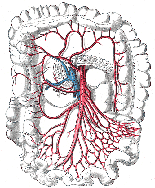
Jejunal and ileal branches of the superior mesenteric artery forming arcades and vasa recta; the terminal ileum also from the ileocolic artery.
- Venous drainage
Superior mesenteric vein → hepatic portal vein.
- Lymphatic drainage
Lacteals → mesenteric nodes → superior mesenteric nodes → intestinal trunk → cisterna chyli.
- Innervation
Vagus (parasympathetic) and sympathetic (about T8–T10 via the superior mesenteric plexus). Visceral pain refers to the periumbilical region (midgut).

Clinical correlations
Small bowel obstruction PANCE
- Mechanism / lesion
Adhesions from prior surgery (most common in adults), hernias, and malignancy; Crohn strictures; gallstone ileus; intussusception in children.
- Classic presentation
Crampy periumbilical pain, distension, vomiting (early with proximal obstruction), and obstipation. High-pitched, tinkling bowel sounds early. Fever, tachycardia, peritonitis or constant pain suggest strangulation.
Meckel diverticulum PANCE
- Mechanism / lesion
Persistent vitelline (omphalomesenteric) duct, a true diverticulum on the antimesenteric border of the ileum. Rule of 2s: about 2% of people, about 2 feet from the ileocecal valve, about 2 inches long, 2 types of ectopic tissue (gastric, pancreatic), symptoms often before age 2.
- Classic presentation
Painless rectal bleeding in a young child (acid from ectopic gastric mucosa ulcerates the adjacent ileum), intussusception, obstruction, or diverticulitis mimicking appendicitis. Technetium-99m pertechnetate scan (Meckel scan) detects the gastric mucosa.
Crohn disease PANCE
- Mechanism / lesion
Transmural granulomatous inflammation that can affect any part of the GI tract but most commonly the terminal ileum, with skip lesions.
- Classic presentation
Chronic diarrhea, RLQ pain, weight loss, fistulas, strictures, perianal disease, B12 deficiency and gallstones (from bile acid loss).
Acute mesenteric ischemia PANCE
- Mechanism / lesion
Most often embolus to the SMA (atrial fibrillation), also thrombosis, non-occlusive ischemia, or venous thrombosis.
- Classic presentation
Pain out of proportion to exam in an older patient with AF or vascular disease, elevated lactate late. CT angiography; emergency revascularization.
Intussusception PANCE
- Mechanism / lesion
Telescoping of bowel (usually ileum into colon, ileocolic), most common at 6–36 months; lead points in older children and adults.
- Classic presentation
Intermittent severe colicky pain with drawing up of the legs, vomiting, "currant jelly" stool (late), and a sausage-shaped mass. Ultrasound shows a target sign. Air or contrast enema is diagnostic and therapeutic.
High-yield PANCE pearls
- Small bowel obstruction #1 cause in adults: adhesions. Look for air-fluid levels and dilated loops.
- Meckel: painless rectal bleeding in a toddler; Tc-99m pertechnetate scan; rule of 2s.
- Crohn: terminal ileum, skip lesions, transmural, string sign; B12 malabsorption.
- Mesenteric ischemia: pain out of proportion + AF → CT angiography.
- Intussusception: currant jelly stool, sausage mass, target sign on ultrasound; enema reduction.
Imaging anatomy
- X-ray
Obstruction: dilated small bowel loops (more than about 3 cm) with air-fluid levels in a step-ladder pattern and little colonic gas. Free air with perforation. verify
- CT
CT with IV contrast is the main test for obstruction (a transition point, closed loop, signs of ischemia), Crohn disease (CT/MR enterography: wall thickening, "comb sign", fistulas), and CT angiography for mesenteric ischemia.
- MRI
MR enterography for Crohn disease activity and complications without radiation.
- Ultrasound
First-line for intussusception in children (target or doughnut sign). Can show dilated fluid-filled loops and bowel wall thickening.
- Key views & landmarks
- Supine and upright abdominal radiographs
- CT abdomen/pelvis with contrast
- Tc-99m pertechnetate scan for Meckel
Physical exam
- Surface landmarks
Jejunum in the left upper and central abdomen; terminal ileum in the right iliac fossa.
- Palpation
Distension, tenderness, masses (RLQ mass in Crohn disease; sausage mass in intussusception). Check hernia orifices in every obstruction.
- Strength / function testing
Auscultate for bowel sounds (high-pitched and tinkling early in obstruction; reduced or absent in ileus or late obstruction). Percuss for tympany.
Self-quiz
A 2-year-old boy has painless passage of a large amount of bright red and maroon blood per rectum. He is otherwise well. Which test is most likely to confirm the diagnosis?
Correct answer: B
Meckel diverticulum with ectopic gastric mucosa causes painless bleeding in young children. A Tc-99m pertechnetate (Meckel) scan detects the gastric mucosa.
Show answer
B
Meckel diverticulum with ectopic gastric mucosa causes painless bleeding in young children. A Tc-99m pertechnetate (Meckel) scan detects the gastric mucosa.
A 60-year-old woman with prior hysterectomy has crampy abdominal pain, vomiting, distension and no flatus for 24 hours. What is the most likely cause?
Correct answer: B
Adhesions from prior abdominal or pelvic surgery are the most common cause of small bowel obstruction in adults.
Show answer
B
Adhesions from prior abdominal or pelvic surgery are the most common cause of small bowel obstruction in adults.
Which feature distinguishes the ileum from the jejunum?
Correct answer: D
The ileum has many arcades, short vasa recta, more fat and Peyer patches. The jejunum has long vasa recta, few arcades, prominent folds and a thicker wall.
Show answer
D
The ileum has many arcades, short vasa recta, more fat and Peyer patches. The jejunum has long vasa recta, few arcades, prominent folds and a thicker wall.
A 78-year-old man with atrial fibrillation has sudden severe abdominal pain, but his abdomen is soft with only mild tenderness. What is the most appropriate next test?
Correct answer: B
Pain out of proportion to exam in a patient with AF suggests acute mesenteric ischemia from an SMA embolus. CT angiography is the diagnostic test of choice.
Show answer
B
Pain out of proportion to exam in a patient with AF suggests acute mesenteric ischemia from an SMA embolus. CT angiography is the diagnostic test of choice.
A 14-month-old has episodes of severe colicky pain with drawing up of the legs and a sausage-shaped abdominal mass. Ultrasound shows a target sign. What is the most appropriate treatment for an otherwise stable child?
Correct answer: D
Intussusception (usually ileocolic) is diagnosed by ultrasound (target sign), and an air or contrast enema is both diagnostic and therapeutic in a stable child. Surgery is reserved for failed reduction, perforation or peritonitis.
Show answer
D
Intussusception (usually ileocolic) is diagnosed by ultrasound (target sign), and an air or contrast enema is both diagnostic and therapeutic in a stable child. Surgery is reserved for failed reduction, perforation or peritonitis.