Lumbar plexus
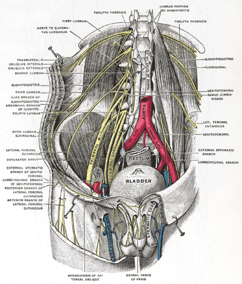
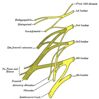
Nerve network formed within psoas major from the anterior rami of L1–L4. It supplies the lower abdominal wall, groin and anterior and medial thigh, and its branches are injured in hernia repair and by tight clothing.
Core anatomy
- Location
Within the substance of psoas major, on the posterior abdominal wall.
- Root values
Anterior rami of L1–L4 (often with a contribution from T12).
- Origin
Anterior rami of L1–L4 in psoas major.
- Course
Branches emerge from the lateral border, medial border or anterior surface of psoas and pass to the abdominal wall, groin and thigh.
- Branches
- Iliohypogastric nerve (L1): skin over the iliac crest and hypogastrium; internal oblique and transversus
- Ilioinguinal nerve (L1): through the inguinal canal; skin of the medial thigh, root of the penis and anterior scrotum or mons pubis and labia majora
- Genitofemoral nerve (L1–L2): genital branch (cremaster, scrotal skin) and femoral branch (skin over the femoral triangle)
- Lateral femoral cutaneous nerve (L2–L3): skin of the lateral thigh; passes under or through the inguinal ligament near the ASIS
- Femoral nerve (L2–L4, posterior divisions): anterior thigh muscles (quadriceps, sartorius, pectineus, with iliacus); skin of the anterior thigh and medial leg (saphenous nerve)
- Obturator nerve (L2–L4, anterior divisions): medial thigh adductors; skin of the medial thigh
- Lumbosacral trunk (L4–L5): joins the sacral plexus
- Motor
Lower abdominal wall muscles (iliohypogastric, ilioinguinal), cremaster, hip flexors, knee extensors (femoral), and hip adductors (obturator).
- Sensory
Lower abdominal wall, groin, genitalia (anterior), and the anterior, lateral and medial thigh; medial leg (saphenous).
- Notes
Mnemonic for the branches from superior to inferior: "I I Get Lunch On Fridays" (Iliohypogastric, Ilioinguinal, Genitofemoral, Lateral femoral cutaneous, Obturator, Femoral).
Muscles innervated
- Psoas major — Anterior rami of lumbar nerves L1–L3 (from the lumbar plexus)
Muscles innervated via branches
- Adductor muscles of the thigh — via Femoral nerve
- Quadriceps femoris — via Femoral nerve
- Adductor muscles of the thigh — via Obturator nerve
Branches
Other structures innervated
Mentioned in
Generated automatically from links in other entries.


Clinical correlations
Meralgia paresthetica PANCE
- Mechanism / lesion
Compression of the lateral femoral cutaneous nerve as it passes under the inguinal ligament near the ASIS (obesity, pregnancy, tight belts or pants, diabetes).
- Classic presentation
Burning, tingling or numbness of the anterolateral thigh, no motor weakness, normal reflexes. Usually managed conservatively (loosen clothing, weight loss).
Ilioinguinal and iliohypogastric nerve injury
- Mechanism / lesion
Entrapment or injury during inguinal hernia repair, Pfannenstiel (low transverse) incisions, or laparoscopic port placement.
- Classic presentation
Chronic groin pain or numbness over the groin, medial thigh, and scrotum or labia.
Femoral and obturator nerve injury PANCE
- Mechanism / lesion
Femoral: psoas/retroperitoneal hematoma, pelvic surgery (retractor compression), hip surgery. Obturator: pelvic surgery or tumor, childbirth.
- Classic presentation
Femoral: weak knee extension (knee buckling), reduced patellar reflex (L4), numbness of the anterior thigh and medial leg. Obturator: weak thigh adduction and a wide-based gait, numbness of the medial thigh.
High-yield PANCE pearls
- Meralgia paresthetica = lateral femoral cutaneous nerve; lateral thigh burning, no weakness.
- Femoral nerve (L2–L4): knee extension + patellar reflex (L4).
- Obturator nerve: thigh adduction.
- Cremasteric reflex: L1–L2 (genitofemoral).
Imaging anatomy
- CT
CT shows retroperitoneal hematoma or masses compressing plexus branches.
- MRI
MR neurography for plexopathy (tumor, inflammation); MRI of the lumbar spine distinguishes radiculopathy from plexopathy.
- Ultrasound
Identifies the lateral femoral cutaneous nerve near the ASIS and the femoral nerve lateral to the femoral artery; used for nerve blocks.
- Key views & landmarks
- EMG/nerve conduction studies to localize (root vs plexus vs peripheral nerve)
Physical exam
- Surface landmarks
The lateral femoral cutaneous nerve passes about 1 cm medial to the ASIS under the inguinal ligament. The femoral nerve lies lateral to the femoral artery in the femoral triangle ("NAVEL", lateral to medial).
- Strength / function testing
Hip flexion (L2–L3), knee extension (L3–L4), hip adduction (obturator), patellar reflex (L4), cremasteric reflex (L1–L2), and sensory mapping of the thigh.
Self-quiz
A 32-year-old pregnant woman has burning and numbness of the anterolateral thigh without weakness. Reflexes are normal. Which nerve is affected?
Correct answer: C
Meralgia paresthetica is compression of the purely sensory lateral femoral cutaneous nerve under the inguinal ligament, common with pregnancy, obesity and tight clothing.
Show answer
C
Meralgia paresthetica is compression of the purely sensory lateral femoral cutaneous nerve under the inguinal ligament, common with pregnancy, obesity and tight clothing.
After a pelvic operation, a patient has difficulty climbing stairs, a buckling knee and an absent patellar reflex. Which nerve is injured?
Correct answer: B
The femoral nerve (L2–L4) supplies the quadriceps; injury weakens knee extension and reduces the patellar reflex.
Show answer
B
The femoral nerve (L2–L4) supplies the quadriceps; injury weakens knee extension and reduces the patellar reflex.
Which roots form the lumbar plexus?
Correct answer: B
The lumbar plexus forms within psoas major from the anterior rami of L1–L4, often with a T12 contribution. L4–S3 form the sacral plexus.
Show answer
B
The lumbar plexus forms within psoas major from the anterior rami of L1–L4, often with a T12 contribution. L4–S3 form the sacral plexus.