Cervical plexus
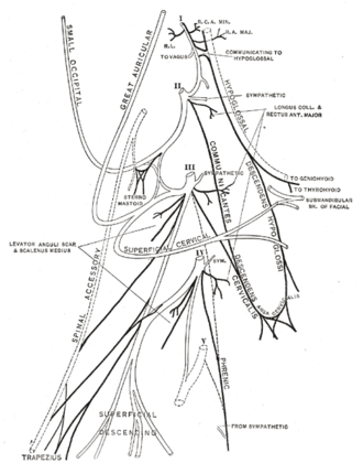
Network of the C1–C4 ventral rami deep to sternocleidomastoid. It gives cutaneous branches to the neck and ear, the ansa cervicalis to the strap muscles, and most importantly the phrenic nerve (C3–C5) to the diaphragm.
Core anatomy
- Root values
C1–C4 ventral rami (phrenic nerve C3–C5)
- Origin
Ventral rami of C1–C4, forming loops in front of levator scapulae and middle scalene, deep to sternocleidomastoid and the internal jugular vein.
- Course
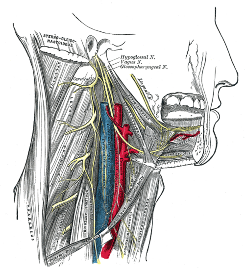
The cutaneous branches emerge around the midpoint of the posterior border of sternocleidomastoid (the nerve point of the neck, Erb point) and fan out over the neck, ear, and shoulder. Motor branches pass to deep neck muscles and, via the ansa cervicalis on the carotid sheath, to the infrahyoid muscles.
- Branches
- Lesser occipital (C2) — skin behind the ear
- Great auricular (C2–C3) — skin over the parotid, angle of the jaw and auricle (at risk in parotid surgery; see parotid gland)
- Transverse cervical (C2–C3) — skin of the anterior neck
- Supraclavicular nerves (C3–C4) — skin over the clavicle and top of the shoulder (referred shoulder pain)
- Ansa cervicalis (C1–C3) — superior root (C1, running with the hypoglossal nerve) and inferior root (C2–C3); supplies the infrahyoid (strap) muscles — sternohyoid, sternothyroid, omohyoid (C1 fibers via CN XII also reach thyrohyoid and geniohyoid)
- Phrenic nerve (C3–C5) — motor to the diaphragm; sensory to the central diaphragm, pericardium and mediastinal pleura
- Branches to SCM and trapezius (C2–C4, proprioceptive), prevertebral muscles, levator scapulae and scalenes
- Motor
Infrahyoid muscles (ansa cervicalis), the diaphragm (phrenic), prevertebral and scalene muscles.
- Sensory
Skin of the anterolateral neck, the angle of the jaw and parotid region, the lower auricle and back of the ear, and over the clavicle and shoulder. (The face above is trigeminal; the back of the scalp is the greater occipital nerve, a dorsal ramus.)
- Relations
Lies on the prevertebral fascia deep to the carotid sheath; the phrenic nerve descends on the anterior scalene. The spinal accessory nerve emerges near the nerve point of the neck.
- Notes
"C3, 4, 5 keeps the diaphragm alive."
Muscles innervated
- Sternocleidomastoid — Spinal accessory nerve (CN XI) (motor); C2–C3 ventral rami (proprioception, via the cervical plexus).
Muscles innervated via branches
- Diaphragm — via Phrenic nerve
Branches
Other structures innervated
Found in
Mentioned in
Generated automatically from links in other entries.


Clinical correlations
Phrenic nerve and high cervical spinal cord injury PANCE
- Mechanism / lesion
Cord injury at or above C3–C5 disrupts phrenic output.
- Classic presentation
Injury above C3 → apnea and ventilator dependence; C4–C5 → weakened diaphragmatic breathing. Unilateral phrenic palsy (surgery, tumor, interscalene block) → elevated hemidiaphragm on chest X-ray and paradoxical movement on fluoroscopic sniff test.
Referred shoulder pain PANCE
- Mechanism / lesion
Diaphragmatic or peritoneal irritation (phrenic, C3–C5) is felt in the C3–C4 dermatome — the supraclavicular nerve territory over the shoulder.
- Classic presentation
Kehr sign (left shoulder pain with splenic rupture), right shoulder pain with gallbladder disease or subphrenic abscess, shoulder pain after laparoscopy.
Superficial cervical plexus block and nerve injury
- Mechanism / lesion
Local anesthetic injected at the midpoint of the posterior border of SCM anesthetizes the cutaneous branches (carotid, thyroid, clavicle surgery). Interscalene brachial plexus blocks almost always cause temporary ipsilateral phrenic palsy.
- Classic presentation
Great auricular nerve injury after parotidectomy or facelift → numbness of the earlobe and angle of the jaw.
High-yield PANCE pearls
- "C3, 4, 5 keeps the diaphragm alive" — phrenic nerve.
- Spinal cord injury above C3 → ventilator dependence.
- Shoulder-tip pain from diaphragm irritation = referred via phrenic (C3–C5) → Kehr sign.
- Elevated hemidiaphragm → phrenic nerve palsy → sniff test.
- Ansa cervicalis → strap muscles.
Imaging anatomy
- X-ray
Chest X-ray shows an elevated hemidiaphragm in phrenic palsy.
- MRI
Cervical spinal cord injury level.
- Ultrasound
Diaphragm ultrasound (or fluoroscopic sniff test) shows reduced or paradoxical movement; ultrasound also guides cervical plexus blocks.
Physical exam
- Surface landmarks
Nerve point of the neck — midpoint of the posterior border of SCM.
- Strength / function testing
Sensation over the angle of the jaw and earlobe (great auricular, C2–C3), the anterior neck and over the clavicle (C3–C4); watch diaphragmatic breathing and look for paradoxical abdominal movement in suspected phrenic palsy.
Self-quiz
A 22-year-old has a spinal cord transection at C2 after a diving accident. Which consequence is most expected?
Correct answer: B
The phrenic nerve arises from C3–C5. A complete lesion at C2 cuts off descending respiratory drive to the phrenic motor neurons, causing apnea and ventilator dependence.
Show answer
B
The phrenic nerve arises from C3–C5. A complete lesion at C2 cuts off descending respiratory drive to the phrenic motor neurons, causing apnea and ventilator dependence.
Twelve hours after laparoscopic cholecystectomy, a patient has pain at the tip of the right shoulder. Which nerve roots explain this referred pain?
Correct answer: B
Irritation of the diaphragm (by residual CO₂ or blood) is carried by the phrenic nerve (C3–C5) and felt in the C3–C4 dermatome over the shoulder (supraclavicular nerves).
Show answer
B
Irritation of the diaphragm (by residual CO₂ or blood) is carried by the phrenic nerve (C3–C5) and felt in the C3–C4 dermatome over the shoulder (supraclavicular nerves).
Where do the cutaneous branches of the cervical plexus emerge (the "nerve point" of the neck)?
Correct answer: C
The lesser occipital, great auricular, transverse cervical and supraclavicular nerves emerge around the midpoint of the posterior border of SCM (the nerve point, Erb point), where a superficial cervical plexus block is placed. The spinal accessory nerve emerges nearby.
Show answer
C
The lesser occipital, great auricular, transverse cervical and supraclavicular nerves emerge around the midpoint of the posterior border of SCM (the nerve point, Erb point), where a superficial cervical plexus block is placed. The spinal accessory nerve emerges nearby.
After a parotidectomy, a patient has numbness of the earlobe and the skin over the angle of the jaw. Which nerve was injured?
Correct answer: E
The great auricular nerve (C2–C3) supplies the skin over the parotid, the angle of the jaw and the lower auricle, and is at risk in parotid surgery and facelifts.
Show answer
E
The great auricular nerve (C2–C3) supplies the skin over the parotid, the angle of the jaw and the lower auricle, and is at risk in parotid surgery and facelifts.
The ansa cervicalis supplies which muscles?
Correct answer: A
The ansa cervicalis (C1–C3), lying on the carotid sheath, supplies the infrahyoid (strap) muscles: sternohyoid, sternothyroid and omohyoid. The diaphragm is supplied by the phrenic nerve (C3–C5).
Show answer
A
The ansa cervicalis (C1–C3), lying on the carotid sheath, supplies the infrahyoid (strap) muscles: sternohyoid, sternothyroid and omohyoid. The diaphragm is supplied by the phrenic nerve (C3–C5).