Ureter
Muscular tube carrying urine from the renal pelvis to the bladder. Its three narrow points are where stones lodge, and its pelvic course puts it at risk during gynecologic surgery.
Core anatomy
- Location
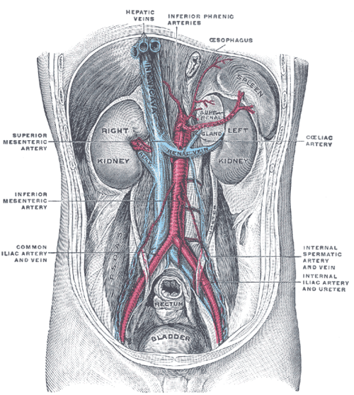
Retroperitoneal, descending on the psoas major from the renal pelvis to the bladder. About 25 cm long.
- Parts
- Abdominal part: from the ureteropelvic junction down along the psoas, crossing the genitofemoral nerve and passing behind the gonadal vessels
- Pelvic part: crosses the pelvic brim at about the bifurcation of the common iliac artery, descends on the lateral pelvic wall, then turns medially to enter the bladder obliquely
- Three physiological constrictions: (1) ureteropelvic junction; (2) where it crosses the iliac vessels / pelvic brim; (3) ureterovesical junction (the narrowest)
- Relations
"Water under the bridge": in females the ureter passes under the uterine artery near the lateral fornix of the cervix; in males it passes under the ductus deferens. The gonadal vessels cross anterior to it in the abdomen.
- Arterial supply
Segmental, from the renal artery (upper), gonadal artery and aorta (middle), and internal iliac branches (lower, e.g. vesical and uterine arteries). Longitudinal anastomoses along the adventitia; stripping it risks ischemia.
- Innervation
Renal, aortic and hypogastric plexuses (about T11–L2). Pain refers from the loin to the groin (and labia or scrotum) as a stone descends.
Mentioned in
- Broad ligament
- Ductus deferens and seminal glands
- Internal iliac artery
- Kidney
- Ovary
- Psoas major
- Sacral plexus
- Urinary bladder
- Uterine artery
- Uterus
- Vagina
Generated automatically from links in other entries.
Swipe sideways to see the whole diagram →

Clinical correlations
Ureteral calculus (renal colic) PANCE
- Mechanism / lesion
Stones lodge at the three narrow points, most often the ureterovesical junction.
- Classic presentation
Colicky flank pain radiating to the groin, testis or labia; hematuria; nausea. Distal stones cause urinary urgency and frequency. Most stones under about 5 mm pass spontaneously; alpha-blockers (tamsulosin) are offered to help pass distal ureteral stones up to 10 mm (AUA). Obstruction with infection, a solitary kidney, AKI or intractable pain needs urgent urology (stent or nephrostomy).
Iatrogenic ureteral injury PANCE
- Mechanism / lesion
Ligation, transection or thermal injury during hysterectomy (near the uterine artery), oophorectomy (near the gonadal vessels at the pelvic brim), or colorectal surgery.
- Classic presentation
Postoperative flank pain, fever, urinoma, hydronephrosis, rising creatinine (if bilateral), or urine leaking from the vagina (ureterovaginal fistula).
Ureteropelvic junction obstruction
- Mechanism / lesion
Congenital narrowing or a crossing lower-pole vessel. The most common cause of prenatal hydronephrosis.
- Classic presentation
Hydronephrosis on prenatal ultrasound; in adults, intermittent flank pain after high fluid intake (e.g. alcohol).
High-yield PANCE pearls
- Stones lodge at the UPJ, pelvic brim (iliac vessels) and UVJ (narrowest).
- Water under the bridge: ureter passes under the uterine artery (female) / ductus deferens (male).
- Hysterectomy is the classic setting for ureteral injury.
- Obstructing stone + fever = emergency decompression.
Imaging anatomy
- X-ray
KUB shows radiopaque stones along the ureteral course (often confused with pelvic phleboliths).
- CT
Non-contrast CT for stones (the "rim sign" of ureteral wall around a stone helps distinguish it from a phlebolith). CT urography for the collecting system and ureteral injury (contrast leak).
- Ultrasound
Shows hydronephrosis and UVJ stones through a full bladder; ureteral jets on color Doppler. Preferred in pregnancy.
- Key views & landmarks
- Non-contrast CT (stone protocol)
- CT urography (delayed excretory phase)
Physical exam
- Surface landmarks
The ureter runs roughly along the tips of the lumbar transverse processes on radiographs and crosses the pelvic brim near the sacroiliac joint.
- Strength / function testing
CVA tenderness and suprapubic tenderness. Pain migrating from the flank to the groin suggests a descending stone.
Self-quiz
Where does a ureteral stone most commonly become lodged?
Correct answer: D
The ureterovesical junction is the narrowest point of the ureter and the most common site of impaction. The UPJ and pelvic brim are the other constrictions.
Show answer
D
The ureterovesical junction is the narrowest point of the ureter and the most common site of impaction. The UPJ and pelvic brim are the other constrictions.
During a hysterectomy, the ureter is at greatest risk of injury where it passes beneath which structure?
Correct answer: B
The ureter passes under the uterine artery ("water under the bridge") about 1–2 cm lateral to the cervix, where it can be clamped or ligated during hysterectomy.
Show answer
B
The ureter passes under the uterine artery ("water under the bridge") about 1–2 cm lateral to the cervix, where it can be clamped or ligated during hysterectomy.
A 40-year-old man with a 6-mm distal ureteral stone has a temperature of 39.2 °C and hydronephrosis. What is the most appropriate management?
Correct answer: B
An infected obstructed kidney is a urologic emergency requiring urgent decompression and IV antibiotics. Medical expulsive therapy is only for uncomplicated stones.
Show answer
B
An infected obstructed kidney is a urologic emergency requiring urgent decompression and IV antibiotics. Medical expulsive therapy is only for uncomplicated stones.
At the pelvic brim, the ureter crosses which structure?
Correct answer: D
The ureter enters the pelvis by crossing the bifurcation of the common iliac artery at the pelvic brim, one of its three narrow points (with the UPJ and UVJ). Lower in the pelvis it passes under the uterine artery or ductus deferens.
Show answer
D
The ureter enters the pelvis by crossing the bifurcation of the common iliac artery at the pelvic brim, one of its three narrow points (with the UPJ and UVJ). Lower in the pelvis it passes under the uterine artery or ductus deferens.
What is the most common cause of hydronephrosis detected on prenatal ultrasound?
Correct answer: B
Ureteropelvic junction obstruction (congenital narrowing or a crossing lower-pole vessel) is the most common cause of prenatal hydronephrosis. In adults it can cause intermittent flank pain after high fluid intake.
Show answer
B
Ureteropelvic junction obstruction (congenital narrowing or a crossing lower-pole vessel) is the most common cause of prenatal hydronephrosis. In adults it can cause intermittent flank pain after high fluid intake.