Rectum
Final part of the large intestine, from the rectosigmoid junction at S3 to the anorectal junction at the puborectalis sling. Rectal cancer staging and treatment depend on its peritoneal relations and pelvic surroundings.
Core anatomy
- Location
Follows the concavity of the sacrum and coccyx and coccyx in the posterior lesser pelvis, from the rectosigmoid junction (about S3) to the anorectal junction, where it passes through the pelvic diaphragm and becomes the anal canal. About 12–15 cm long.
- Parts
- Rectosigmoid junction: where the taeniae coli spread out into a continuous outer longitudinal layer and the sigmoid mesocolon ends; the rectum has no taeniae, haustra, epiploic appendices or mesentery
- Sacral flexure following the sacrum; anorectal flexure (perineal flexure, about 80°) made by the puborectalis sling
- Transverse rectal folds (valves of Houston): usually three, two on the left and one (the largest, middle) on the right
- Rectal ampulla: the dilated lower part that stores feces
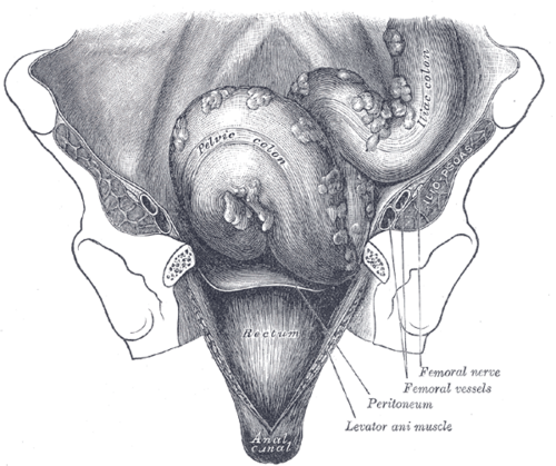
- Peritoneum: covers the front and sides of the upper third, the front only of the middle third; the lower third is below the peritoneum. Reflects onto the bladder (rectovesical pouch) or posterior vaginal fornix and uterus (rectouterine pouch)
- Relations
Posterior: sacrum, coccyx, the sacral plexus and median sacral vessels. Anterior — male: Urinary bladder, seminal glands, ductus deferens and seminal glands and prostate (behind the rectoprostatic fascia). Female: Vagina, cervix and the rectouterine pouch. The mesorectum is the fatty sheath containing its vessels and nodes.
- Arterial supply
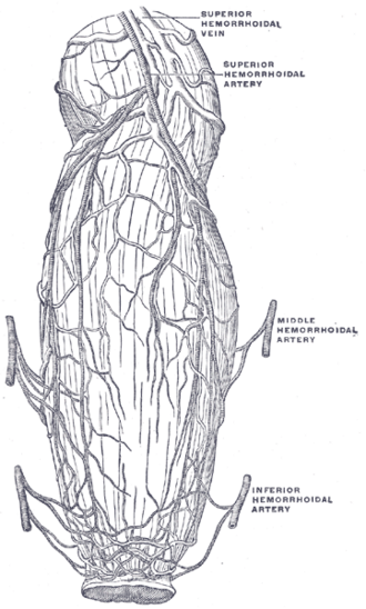
Superior rectal artery (continuation of the inferior mesenteric artery) — the main supply; middle rectal arteries (from the internal iliac artery); inferior rectal arteries (from the internal pudendal) to the anorectal junction.
- Venous drainage
Superior rectal vein → inferior mesenteric vein → portal system; middle and inferior rectal veins → internal iliac/internal pudendal veins (systemic). The rectal venous plexus is a portosystemic anastomosis.
- Lymphatic drainage
Upper rectum → pararectal and inferior mesenteric nodes (along the superior rectal vessels); lower rectum → internal iliac and sacral nodes (see pelvic and inguinal lymph drainage).
- Innervation
Sympathetic from the lumbar splanchnic nerves via the hypogastric plexuses; parasympathetic from the pelvic splanchnic nerves (S2–S4) (see pelvic autonomic nerves). Visceral afferents carry the sensation of rectal fullness.
Mentioned in
- Anal canal
- Bony pelvis
- Ductus deferens and seminal glands
- Internal iliac artery
- Pelvic and inguinal lymph drainage
- Pelvic autonomic nerves
- Prostate
- Rectouterine pouch
- Urinary bladder
- Uterus
- Vagina
Generated automatically from links in other entries.


Clinical correlations
Rectal cancer PANCE
- Mechanism / lesion
Adenocarcinoma arising from adenomatous polyps; risk factors as for colorectal cancer (age, family history, IBD, Lynch syndrome, FAP).
- Classic presentation
Hematochezia, change in bowel habit, narrow stools, tenesmus, iron-deficiency anemia; a mass may be felt on DRE.
- Notes
Local staging with pelvic MRI (depth, nodes, circumferential resection margin) or endorectal ultrasound. Locally advanced tumors get neoadjuvant chemoradiation, then total mesorectal excision. Screening for average-risk adults begins at 45 (USPSTF 2021).
Rectal prolapse
- Mechanism / lesion
Full-thickness protrusion of the rectal wall through the anus; associated with chronic straining, pelvic floor weakness, and cystic fibrosis in children.
- Classic presentation
A protruding mass with concentric mucosal folds (hemorrhoids show radial folds), mucus discharge, incontinence.
Proctitis
- Mechanism / lesion
Inflammation of the rectal mucosa — ulcerative colitis (always starts in the rectum), STIs (gonorrhea, chlamydia including LGV, HSV, syphilis), radiation.
- Classic presentation
Tenesmus, urgency, rectal bleeding and mucus.
High-yield PANCE pearls
- Ulcerative colitis always involves the rectum and extends proximally.
- Rectal cancer local staging = pelvic MRI; surgery = total mesorectal excision.
- CRC screening starts at 45 for average risk.
- Rectal prolapse = concentric rings; prolapsed hemorrhoids = radial folds.
- Rectal venous plexus = portosystemic anastomosis (portal hypertension).
Imaging anatomy
- CT
CT chest/abdomen/pelvis for distant staging (liver and lung metastases).
- MRI
High-resolution pelvic MRI is the key local staging study: tumor depth, mesorectal fascia involvement (predicts the circumferential resection margin), extramural vascular invasion and nodes.
- Ultrasound
Endorectal ultrasound stages early (T1–T2) tumors.
- Key views & landmarks
- Pelvic MRI rectal cancer protocol
- Colonoscopy with biopsy
Physical exam
- Surface landmarks
The lower rectum is within reach of the examining finger (about 7–8 cm from the anal verge); the prostate or cervix is felt anteriorly.
- Palpation
DRE: note masses (distance from the anal verge, fixed or mobile), tenderness, stool character, and blood on the glove. An anterior shelf-like mass can indicate peritoneal metastases in the rectovesical/rectouterine pouch (Blumer shelf).
- Strength / function testing
Stool guaiac/FIT is for screening, not for evaluating a patient who has symptoms.
Self-quiz
A 61-year-old man has 3 months of rectal bleeding, tenesmus and thinner stools. A mass is felt 6 cm from the anal verge and biopsy shows adenocarcinoma. What is the best study for local staging?
Correct answer: B
Pelvic MRI defines tumor depth, nodal involvement and the relationship to the mesorectal fascia, which guides neoadjuvant therapy and total mesorectal excision. CT staging is for distant spread.
Show answer
B
Pelvic MRI defines tumor depth, nodal involvement and the relationship to the mesorectal fascia, which guides neoadjuvant therapy and total mesorectal excision. CT staging is for distant spread.
The superior rectal artery is a continuation of which artery?
Correct answer: C
The superior rectal artery is the terminal continuation of the inferior mesenteric artery, and it is the main supply to the rectum. The middle rectal arteries come from the internal iliac and the inferior rectal from the internal pudendal.
Show answer
C
The superior rectal artery is the terminal continuation of the inferior mesenteric artery, and it is the main supply to the rectum. The middle rectal arteries come from the internal iliac and the inferior rectal from the internal pudendal.
Which feature distinguishes the rectum from the sigmoid colon?
Correct answer: C
At the rectosigmoid junction the taeniae coli spread out into a continuous longitudinal layer; the rectum has no taeniae, haustra, epiploic appendices or mesentery.
Show answer
C
At the rectosigmoid junction the taeniae coli spread out into a continuous longitudinal layer; the rectum has no taeniae, haustra, epiploic appendices or mesentery.
A 75-year-old woman has a mass protruding from the anus with concentric mucosal folds, mucus discharge and fecal incontinence. What is the most likely diagnosis?
Correct answer: D
Rectal prolapse is a full-thickness protrusion with concentric mucosal folds; prolapsed hemorrhoids show radial folds. It is associated with chronic straining and pelvic floor weakness.
Show answer
D
Rectal prolapse is a full-thickness protrusion with concentric mucosal folds; prolapsed hemorrhoids show radial folds. It is associated with chronic straining and pelvic floor weakness.
Why is the rectal venous plexus a site of portosystemic anastomosis?
Correct answer: B
The superior rectal vein → inferior mesenteric vein → portal system, whereas the middle and inferior rectal veins → internal iliac/internal pudendal veins (systemic). The plexus joining them is a portosystemic anastomosis that can dilate in portal hypertension.
Show answer
B
The superior rectal vein → inferior mesenteric vein → portal system, whereas the middle and inferior rectal veins → internal iliac/internal pudendal veins (systemic). The plexus joining them is a portosystemic anastomosis that can dilate in portal hypertension.
Flashcards
See also
- Anal canal
- Colon
- Rectouterine pouch
- Pelvic diaphragm
- Foregut, midgut and hindgut (comparison table)
- Lymph drainage of the pelvis and perineum (comparison table)