Broad ligament
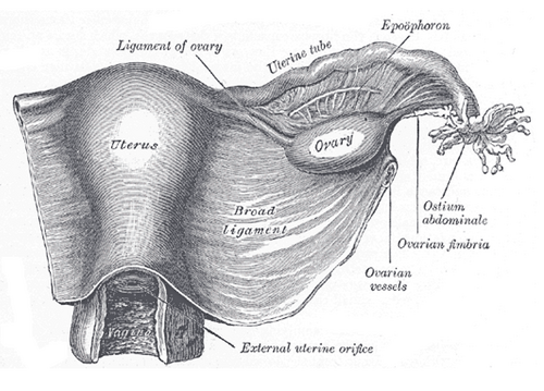
Double fold of peritoneum draped over the uterus, tubes and ovaries to the pelvic walls. It carries their vessels, and the ureter runs beneath its base, where it is at risk during hysterectomy.
Core anatomy
- Location
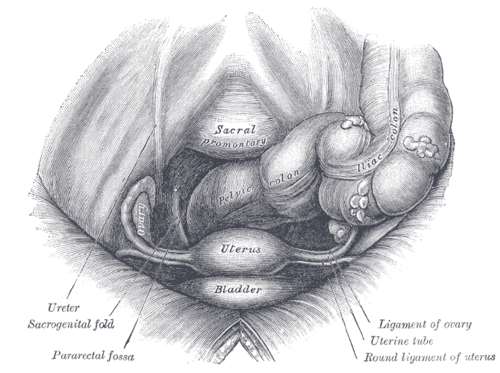
Extends from each side of the uterus to the lateral pelvic wall and floor, dividing the female pelvic cavity into the anterior vesicouterine pouch and the posterior rectouterine pouch.
- Attachments
Medially: the lateral margin of the uterus, where its two layers become the perimetrium. Laterally and inferiorly: the peritoneum of the pelvic wall and floor. Its upper free border encloses the uterine tube; laterally, above the tube's end, it continues as the suspensory ligament of the ovary.
- Function
A mesentery for the uterus, tubes and ovaries (carrying their vessels, nerves and lymphatics); it provides little mechanical support.
- Relations
Parts: mesometrium (the largest part, beside the uterus), mesosalpinx (around the tube) and mesovarium (attaching the ovary). Between its layers: the uterine tube, the round ligament of the uterus and the ligament of the ovary, the uterine artery and uterine venous plexus at its base, the ovarian vessels (laterally, in the suspensory ligament), nerves, lymphatics, and mesonephric remnants (epoophoron, paroophoron). The ureter runs forward under the base, beneath the uterine artery, about 1–2 cm lateral to the cervix.
- Notes
The dense connective tissue at the base of the broad ligament around the uterine vessels forms the cardinal (transverse cervical) ligaments, a major uterine support.
Mentioned in
Generated automatically from links in other entries.


Clinical correlations
Ureteral injury during hysterectomy PANCE
- Mechanism / lesion
Clamping the uterine vessels at the base of the broad ligament, or the ovarian vessels in the suspensory ligament near the pelvic brim, can include the ureter.
- Classic presentation
Postoperative flank pain, fever, urinoma, hydronephrosis or a ureterovaginal fistula (continuous vaginal leakage of urine).
Broad ligament hematoma
- Mechanism / lesion
Bleeding from uterine vessels after delivery (e.g. a cervical or lower-segment laceration extending laterally) or surgery.
- Classic presentation
Pelvic pain, a paravaginal/adnexal mass and signs of hypovolemia despite little external bleeding.
Round ligament pain
- Mechanism / lesion
Stretching of the round ligament (within the broad ligament) as the uterus grows in the second trimester.
- Classic presentation
Sharp, brief groin or lower-quadrant pain with sudden movement; benign.
High-yield PANCE pearls
- Water under the bridge: the ureter passes under the uterine artery at the base of the broad ligament.
- Parts: mesometrium, mesosalpinx, mesovarium.
- Round ligament pain = sharp groin pain in the 2nd trimester with movement; benign.
Imaging anatomy
- MRI
Shows broad-ligament fibroids, hematomas and the course of the ureters and vessels.
- Ultrasound
Not seen as a separate structure unless outlined by fluid; paraovarian (paratubal) cysts and broad ligament fibroids appear separate from the ovary.
Physical exam
- Surface landmarks
Not directly palpable. A mass lateral to the uterus may lie within the broad ligament (fibroid, hematoma, paraovarian cyst).
Self-quiz
During an abdominal hysterectomy, the uterine artery is ligated at the base of the broad ligament. Which structure lies immediately beneath it and is at greatest risk?
Correct answer: B
The ureter runs beneath the uterine artery ("water under the bridge") about 1–2 cm lateral to the cervix at the base of the broad ligament.
Show answer
B
The ureter runs beneath the uterine artery ("water under the bridge") about 1–2 cm lateral to the cervix at the base of the broad ligament.
Which part of the broad ligament encloses the uterine tube?
Correct answer: B
The mesosalpinx is the part of the broad ligament around the uterine tube; the mesovarium attaches the ovary and the mesometrium lies beside the uterus.
Show answer
B
The mesosalpinx is the part of the broad ligament around the uterine tube; the mesovarium attaches the ovary and the mesometrium lies beside the uterus.
Which structure provides major support to the uterus at the base of the broad ligament?
Correct answer: A
The dense connective tissue around the uterine vessels at the base of the broad ligament forms the cardinal (transverse cervical) ligaments, a major uterine support. The broad ligament itself is a mesentery and gives little mechanical support.
Show answer
A
The dense connective tissue around the uterine vessels at the base of the broad ligament forms the cardinal (transverse cervical) ligaments, a major uterine support. The broad ligament itself is a mesentery and gives little mechanical support.