Deep neck spaces
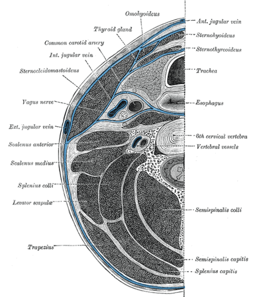
Potential spaces between the layers of deep cervical fascia. Infections can track through them from the teeth, tonsils and pharynx into the mediastinum.
Core anatomy
- Location
Between the layers of the deep cervical fascia, from the skull base into the chest.
- Boundaries
- investing layer
- Surrounds the neck, enclosing sternocleidomastoid, trapezius, and the submandibular and parotid glands
- pretracheal layer
- Encloses the thyroid gland, larynx, trachea, esophagus and infrahyoid muscles (visceral layer includes the buccopharyngeal fascia behind the pharynx)
- prevertebral layer
- Surrounds the vertebral column and deep muscles; its alar part lies in front
- carotid sheath
- Formed from all three layers; contains the common/internal carotid, internal jugular vein and vagus
- Contents
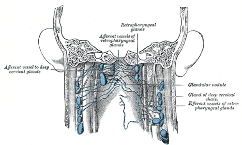
- Retropharyngeal space: between the buccopharyngeal fascia (in front) and the alar fascia (behind), from the skull base to about T1–T2 (upper mediastinum); contains retropharyngeal nodes (prominent in young children) verify
- Danger space: between the alar and prevertebral fascia, from the skull base down to the diaphragm — infection can spread into the posterior mediastinum
- Prevertebral space: deep to the prevertebral fascia along the whole spine (vertebral osteomyelitis, Pott disease)
- Parapharyngeal (lateral pharyngeal) space: inverted pyramid beside the pharynx from the skull base to the hyoid; connects with the other spaces; carotid sheath in its post-styloid part
- Submandibular space: sublingual (above mylohyoid) and submaxillary (below mylohyoid) parts, connected around the back edge of mylohyoid
- Communications
The spaces communicate, so infection from teeth (submandibular), tonsils (peritonsillar → parapharyngeal) and the pharynx (retropharyngeal) can spread into the carotid sheath and down to the mediastinum.
- Notes
Retropharyngeal nodes usually regress by about age 5–6, which is why retropharyngeal abscess is mainly a disease of young children.


Clinical correlations
Retropharyngeal abscess PANCE
- Mechanism / lesion
Suppurative retropharyngeal nodes after URI/pharyngitis in children under 5; in adults, penetrating trauma (fish bone, instrumentation).
- Classic presentation
Fever, sore throat, neck stiffness or pain on extension (can mimic meningitis), torticollis, drooling, muffled voice, stridor. Lateral neck X-ray: widened prevertebral soft tissue; confirm with contrast CT. Treat with IV antibiotics ± surgical drainage; risk of mediastinitis.
Ludwig angina PANCE
- Mechanism / lesion
Rapidly spreading bilateral cellulitis of the submandibular space, usually from infected lower molars (roots below the mylohyoid line); oral streptococci and anaerobes.
- Classic presentation
Brawny, woody submandibular swelling, elevated and pushed-back tongue, drooling, trismus, dysphagia, fever — airway emergency. Secure the airway (awake fiberoptic intubation or tracheostomy), IV antibiotics, drainage if collection.
Descending necrotizing mediastinitis
- Mechanism / lesion
Spread of a deep neck infection down the retropharyngeal or danger space.
- Classic presentation
Chest pain, dyspnea and sepsis after a neck infection; widened mediastinum; high mortality — urgent CT and surgical drainage.
High-yield PANCE pearls
- Child < 5 with fever, sore throat and pain on neck extension → retropharyngeal abscess → widened prevertebral space.
- Woody submandibular swelling, raised tongue after a dental infection → Ludwig angina → airway first.
- Danger space runs from skull base to diaphragm → mediastinitis.
- Deep neck infections → contrast CT of the neck once the airway is secure.
Imaging anatomy
- X-ray
Lateral soft-tissue neck in extension: prevertebral soft tissue wider than about 7 mm at C2 (or more than half the vertebral body width) in children suggests retropharyngeal abscess. verify
- CT
Contrast CT of the neck (± chest) — defines the space involved, distinguishes abscess (rim-enhancing collection) from cellulitis, and detects mediastinal spread.
- MRI
Better soft-tissue detail when needed (e.g. prevertebral/spinal involvement).
- Key views & landmarks
- Lateral neck film
- Contrast CT neck and chest
Physical exam
- Surface landmarks
Submandibular region below the mandible; posterior pharyngeal wall (visible bulge in some retropharyngeal abscesses).
- Palpation
Palpate the floor of the mouth and submandibular region (woody induration in Ludwig angina) and the neck for tenderness and fluctuance.
- Strength / function testing
Assess the airway first (stridor, drooling, voice, positioning); neck range of motion (limited extension in retropharyngeal abscess); trismus; teeth for a source.
Self-quiz
A 3-year-old has had fever and sore throat for 3 days. He refuses to extend his neck, is drooling and has a muffled voice. A lateral neck X-ray shows widening of the prevertebral soft tissue. What is the most likely diagnosis?
Correct answer: B
Retropharyngeal abscess — young child, fever, reluctance to extend the neck, drooling, and widened prevertebral soft tissue. Confirm with contrast CT; the danger space can carry infection to the mediastinum.
Show answer
B
Retropharyngeal abscess — young child, fever, reluctance to extend the neck, drooling, and widened prevertebral soft tissue. Confirm with contrast CT; the danger space can carry infection to the mediastinum.
A 45-year-old with a painful lower molar develops fever and a firm, tender swelling under both sides of the jaw. His tongue is pushed up and he is drooling. What is the priority?
Correct answer: B
Ludwig angina — bilateral submandibular space cellulitis from a lower molar — can obstruct the airway quickly. Airway control comes first, then IV antibiotics and drainage.
Show answer
B
Ludwig angina — bilateral submandibular space cellulitis from a lower molar — can obstruct the airway quickly. Airway control comes first, then IV antibiotics and drainage.
Infection in which deep neck space can spread all the way down to the diaphragm?
Correct answer: D
The danger space, between the alar and prevertebral fascia, runs from the skull base down to the diaphragm, so infection can spread into the posterior mediastinum. The retropharyngeal space reaches only the upper mediastinum.
Show answer
D
The danger space, between the alar and prevertebral fascia, runs from the skull base down to the diaphragm, so infection can spread into the posterior mediastinum. The retropharyngeal space reaches only the upper mediastinum.
Ludwig angina most commonly arises from which source?
Correct answer: B
Ludwig angina is a rapidly spreading bilateral cellulitis of the submandibular space, usually from infected lower molars whose roots lie below the mylohyoid line. The airway is the first priority.
Show answer
B
Ludwig angina is a rapidly spreading bilateral cellulitis of the submandibular space, usually from infected lower molars whose roots lie below the mylohyoid line. The airway is the first priority.
Why is retropharyngeal abscess mainly a disease of young children?
Correct answer: E
The retropharyngeal nodes are prominent in young children and usually regress by about age 5–6; suppuration of these nodes after a URI causes the abscess. In adults it follows penetrating trauma (fish bone, instrumentation).
Show answer
E
The retropharyngeal nodes are prominent in young children and usually regress by about age 5–6; suppuration of these nodes after a URI causes the abscess. In adults it follows penetrating trauma (fish bone, instrumentation).